MR Imaging Findings of Tamoxifen-associated Uterine Adenosarcoma: Report of Two Cases
- Affiliations
-
- 1Department of Radiology, Anam Hospital, College of Medicine, Korea University, Seoul, Korea. urorad@korea.ac.kr
- KMID: 2175578
- DOI: http://doi.org/10.13104/imri.2015.19.1.56
Abstract
- Adenosarcoma of the uterus is a rare biphasic tumor containing benign glandular epithelial and malignant mesenchymal components. The tumor has been reported to be associated with antiestrogen therapy, particularly tamoxifen, but there have been a few case reports with MRI. We present two cases of MRI findings of uterine adenosarcoma after antiestrogen therapy, tamoxifen and toremifene in breast cancer patients. The tumor presents as a large polypoid mass occupying the endometrial cavity, and may protrude into the vagina. On MRI, the tumor typically shows solid components with scattered small cysts and heterogeneous enhancement. These findings are not significantly different from conventional adenosarcoma.
Keyword
MeSH Terms
Figure
-
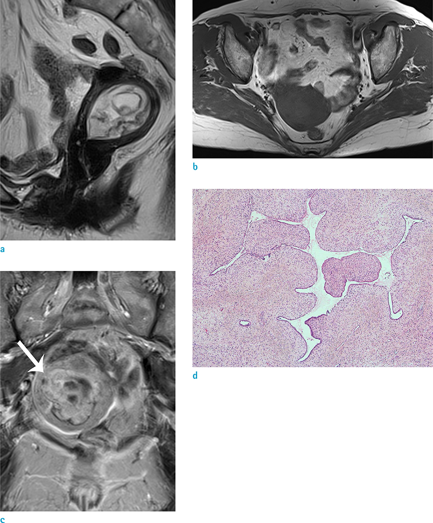
Fig. 1 Case 1. A 69-year-old woman with uterine adenosarcoma. Sagittal turbo spine-echo T2-weighted image (TR/TE, 4000/113) (a) shows the uterine cavity enlarged by a heterogeneous mass which is high signal intensity compared to the myometrium and have well delineated cystic spaces. On axial turbo spin-echo T1-weighted image (TR/TE 538/11) (b), the endometrial mass have intermediate signal intensity. Gadolinium-enhanced turbo spin-echo T1-weighted coronal image (c) shows strong enhancement of the solid components within the tumor. The tumor seemed to invade the myometrium at right side of uterus (arrow). Photomicrograph (× 40) (d) reveals the cellular stroma protruding into a dilated gland lumen. The gland is elongated and compressed, imparting a leaf-like appearance. The stromal cells show high-grade cytologic atypia with mitotic figures (not shown).

Fig. 2 Case 2. A 47-year-old woman with uterine adenosarcoma. Pelvic examination (a) reveals a large polypoid mass protruding to the vagina. Sagittal turbo spine-echo T2-weighted image (TR/TE, 4471/90) (b) shows a heterogeneous polypoid mass occupying the endometrial cavity and protruding from the uterine cervix to the vaginal cavity. The mass shows high signal intensity compared to the myometrium and has tiny hyperintense cystic areas within the mass (arrows). Enlarged uterus with thickened junctional zone is also noted (thick arrows). On axial turbo spin-echo T1-weighted image (TR/TE 550/10) (c), the mass has slightly high signal intensity compared to the myometrium. Tiny scattered high signal intensity areas (arrowheads), presenting hemorrhagic foci are seen. Gadolinium-enhanced fat suppressed turbo spin-echo T1-weighted axial image (d) shows diffuse and strong enhancement of the solid portion of the mass. The mass shows high signal intensity on axial high b-value image (b=1000 s/mm2) (e), but ADC map (f) revealed no diffusion restriction (asterisk). Photograph of gross surgical specimen (g) demonstrates a large yellowish polypoid mass. Small cystic areas scattered within the mass can be observed on the cut surface.
Reference
-
1. Clement PB, Scully RE. Mullerian adenosarcoma of the uterus: a clinicopathologic analysis of 100 cases with a review of the literature. Hum Pathol. 1990; 21:363–381.2. Chourmouzi D, Boulogianni G, Zarampoukas T, Drevelengas A. Sonography and MRI of tamoxifen-associated müllerian adenosarcoma of the uterus. AJR Am J Roentgenol. 2003; 181:1673–1675.3. Yoshizako T, Wada A, Kitagaki H, Ishikawa N, Miyazaki K. MR imaging of uterine adenosarcoma: case report and literature review. Magn Reson Med Sci. 2011; 10:251–254.4. Lee HK, Kim SH, Cho JY, Yeon KM. Uterine adenofibroma and adenosarcoma: CT and MR findings. J Comput Assist Tomogr. 1998; 22:314–316.5. Soh E, Eleti A, Jimenez-Linan M, Arends M, Latimer J, Sala E. Magnetic resonance imaging findings of tamoxifen-associated uterine Mullerian adenosarcoma: a case report. Acta Radiol. 2008; 49:848–851.6. Takeuchi M, Matsuzaki K, Yoshida S, et al. Adenosarcoma of the uterus: magnetic resonance imaging characteristics. Clin Imaging. 2009; 33:244–247.7. Chi F, Wu R, Zeng Y, Xing R, Liu Y, Xu Z. Effects of toremifene versus tamoxifen on breast cancer patients: a meta-analysis. Breast Cancer. 2013; 20:111–122.8. Clement PB. Müllerian adenosarcomas of the uterus with sarcomatous overgrowth. A clinicopathological analysis of 10 cases. Am J Surg Pathol. 1989; 13:28–38.9. Nalaboff KM, Pellerito JS, Ben-Levi E. Imaging the endometrium: disease and normal variants. Radiographics. 2001; 21:1409–1424.10. Shah SH, Jagannathan JP, Krajewski K, O'Regan KN, George S, Ramaiya NH. Uterine sarcomas: then and now. AJR Am J Roentgenol. 2012; 199:213–223.
- Full Text Links
-
- Actions
-
Cited
- CITED
-
- Close
- Share
-
- Similar articles
-
- Endometrial mullerian adenosarcoma after toremifene treatment in breast cancer patients: a case report
- MR Findings of Extrauterine Mullerian Adenosarcoma Associated with Deep Pelvic Endometriosis
- A Case of Mullerian Adenosarcoma of the Uterine Corpus
- A Case of Solitary Brain Metastasis from Uterine Mullerian Adenosarcoma with Sarcomatous Overgrowth
- A Case of Mllerian adenosarcoma of vaginal stump after total abdominal hysterectomy
