A Case of Localized Amyloid Light-Chain Amyloidosis in the Small Intestine
- Affiliations
-
- 1Digestive Disease Center and Research Institute, Department of Internal Medicine, Soonchunhyang University College of Medicine, Bucheon, Korea. kopa9445@schmc.ac.kr
- 2Digestive Disease Center and Research Institute, Department of Pathology, Soonchunhyang University College of Medicine, Bucheon, Korea.
- KMID: 2174384
- DOI: http://doi.org/10.5217/ir.2014.12.3.245
Abstract
- Amyloidosis is characterized by the abnormal deposition of extracellular amyloid fibrils. Cases involving amyloid light-chain amyloidosis in the small intestine have been reported infrequently in Korea. Here, we report a case of localized light chain protein amyloidosis in the small intestine. Esophagogastroduodenoscopy, push enteroscopy, and capsule endoscopy revealed submucosal tumor-like lesions, multiple shallow ulcers, and several erosions in the distal duodenum and jejunum. An endoscopic biopsy established the diagnosis of amyloidosis. In through an immunohistochemical analysis, the presence of lambda light chain protein was detected. The patient had no evidence of an underlying clonal plasma cell disorder or additional organ involvement. Therefore, we concluded that the patient had localized amyloidosis of the small intestine.
MeSH Terms
Figure
-
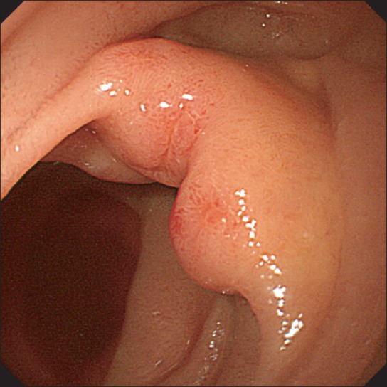
Fig. 1 Upper endoscopic findings. Endoscopy revealed an elevated lesion with central erosion on the third portion of the duodenum.
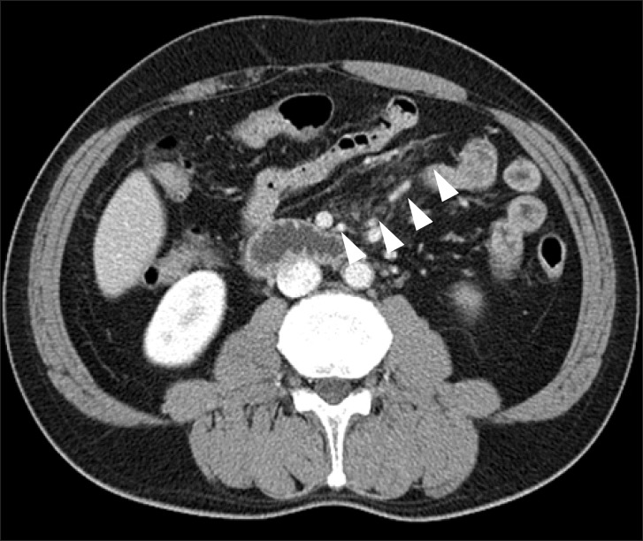
Fig. 2 Abdominal CT findings. Abdominal CT scan revealed haziness in the mesentery (arrows) without abnormal enhancement of wall thickening and dilatation of the small bowel.
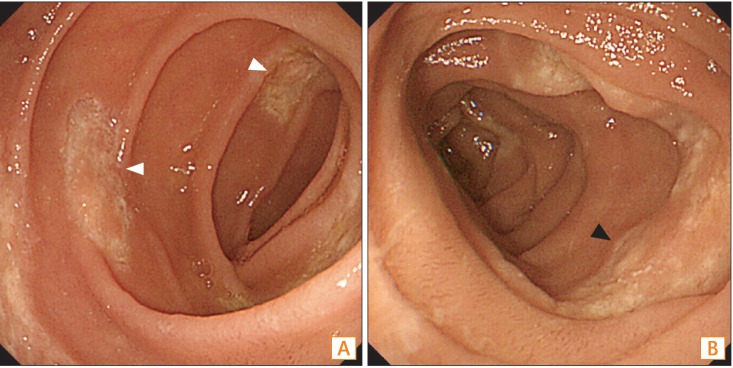
Fig. 3 Push enteroscopic findings. Endoscopy revealed (A) multiple shallow ulcers (white arrows) with broad bases and (B) discoloration of the jejunum (black arrow).
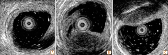
Fig. 4 Endoscopic ultrasonography (EUS) findings. EUS revealed hypoechoic thickening of the muscularis mucosa on (A) a flat lesion with a shallow ulcer and (B) an elevated lesion. Submucosal thickening was present on the elevated lesion (C).
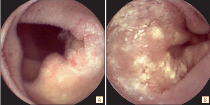
Fig. 5 Capsule endoscopy (CE) findings. CE revealed (A) multiple shallow ulcers and (B) whitish plaque-like infiltration in the jejunum.
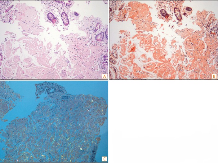
Fig. 6 Histopathologic analysis. Microscopic findings revealed (A) the presence of amorphous eosinophilic material deposits in the lamina propria (×100), (B) salmon-colored deposits of amyloid using Congo red staining (×100), and (C) the apple-green color birefringence of the deposits using polarizing microscopy (×100).
Reference
-
1. Ebert EC, Nagar M. Gastrointestinal manifestations of amyloidosis. Am J Gastroenterol. 2008; 103:776–787. PMID: 18076735.
Article2. Okuda Y, Takasugi K, Oyama T, Onuma M, Oyama H. Amyloidosis in rheumatoid arthritis--clinical study of 124 histologically proven cases. Ryumachi. 1994; 34:939–946. PMID: 7863383.3. Menke DM, Kyle RA, Fleming CR, Wolfe JT 3rd, Kurtin PJ, Oldenburg WA. Symptomatic gastric amyloidosis in patients with primary systemic amyloidosis. Mayo Clin Proc. 1993; 68:763–767. PMID: 8331978.4. Tada S, Iida M, Iwashita A, et al. Endoscopic and biopsy findings of the upper digestive tract in patients with amyloidosis. Gastrointest Endosc. 1990; 36:10–14. PMID: 2311879.5. Park MA, Mueller PS, Kyle RA, Larson DR, Plevak MF, Gertz MA. Primary (AL) hepatic amyloidosis: clinical features and natural history in 98 patients. Medicine (Baltimore). 2003; 82:291–298. PMID: 14530778.6. Tada S, Iida M, Yao T, Kawakubo K, Okada M, Fujishima M. Endoscopic features in amyloidosis of the small intestine: clinical and morphologic differences between chemical types of amyloid protein. Gastrointest Endosc. 1994; 40:45–50. PMID: 8163134.7. Breedveld FC, Markusse HM, MacFarlane JD. Subcutaneous fat biopsy in the diagnosis of amyloidosis secondary to chronic arthritis. Clin Exp Rheumatol. 1989; 7:407–410. PMID: 2591113.8. Grape T, Johansson GW, Eriksson M, Toth E, Thorlacius H. Primary gastroduodenal amyloidosis. Endoscopy. 2011; 43:E288. PMID: 21915830.
Article9. Sawada T, Adachi Y, Akino K, et al. Endoscopic features of primary amyloidosis of the stomach. Endoscopy. 2012; 44(Suppl 2):E275–E276. PMID: 22814919.
Article10. Kim SH, Han JK, Lee KH, et al. Abdominal amyloidosis: spectrum of radiological findings. Clin Radiol. 2003; 58:610–620. PMID: 12887954.
Article11. Araoz PA, Batts KP, MacCarty RL. Amyloidosis of the alimentary canal: radiologic-pathologic correlation of CT findings. Abdom Imaging. 2000; 25:38–44. PMID: 10652919.
Article12. Mekinian A, Jaccard A, Soussan M, et al. 18F-FDG PET/CT in patients with amyloid light-chain amyloidosis: case-series and literature review. Amyloid. 2012; 19:94–98. PMID: 22587492.
Article13. Glaudemans AW, Slart RH, Noordzij W, Dierckx RA, Hazenberg BP. Utility of 18F-FDG PET(/CT) in patients with systemic and localized amyloidosis. Eur J Nucl Med Mol Imaging. 2013; 40:1095–1101. PMID: 23474745.
Article14. Tang YH, Wu YW, Zong J, Wang JC, Miao F. Imaging features of small intestinal amyloidosis: a case report. Abdom Imaging. 2011; 36:694–697. PMID: 21221573.
Article15. Mainenti PP, Segreto S, Mancini M, et al. Intestinal amyloidosis: two cases with different patterns of clinical and imaging presentation. World J Gastroenterol. 2010; 16:2566–2570. PMID: 20503459.
Article16. Kyle RA, Rajkumar SV. Criteria for diagnosis, staging, risk stratification and response assessment of multiple myeloma. Leukemia. 2009; 23:3–9. PMID: 18971951.
Article
- Full Text Links
-
- Actions
-
Cited
- CITED
-
- Close
- Share
-
- Similar articles
-
- Primary Localized Cutaneous Nodular Amyloidosis Following Local Trauma
- Primary Localized Amyloidosis of the Lacrimal Gland
- Primary Localized Cutaneous Nodular Amyloidosis on Scalp Successfully Treated with Excision
- A Case of Nodular Cutaneous Amyloidosis
- Primary Tracheobronchial Amyloidosis: A Case Report
