Ann Dermatol.
2011 Nov;23(4):515-518.
Primary Localized Cutaneous Nodular Amyloidosis Following Local Trauma
- Affiliations
-
- 1Department of Dermatology, School of Medicine and Medical Research Institute, Chungbuk National University, Cheongju, Korea. tyyoon@chungbuk.ac.kr
- 2Department of Internal Medicine, School of Medicine and Medical Research Institute, Chungbuk National University, Cheongju, Korea.
Abstract
- Primary localized cutaneous nodular amyloidosis (nodular amyloidosis) is a rare and distinct type of amyloidosis, in which amyloid L deposition is limited to the skin and typically manifested as a tumefactive nodule on the acral sites. However, the definite cause of nodular amyloidosis is still unknown. Although it is relatively well known that the amyloid deposits in nodular amyloidosis originate from immunoglobulin light chains secreted by local plasma cells, traumatic injury to the skin has rarely been recognized as a triggering factor of nodular amyloidosis. Herein, we present a case of a 50-year-old male patient with primary localized cutaneous nodular amyloidosis, which occurred after local trauma, and discuss the relationship between traumatic damage and dermal amyloid L deposition.
MeSH Terms
Figure
-

Fig. 1 A 5×5 cm sized, well-demarcated, dome-shaped, salmon-colored, waxy nodule with overlying purpuric plaques on the frontal scalp.
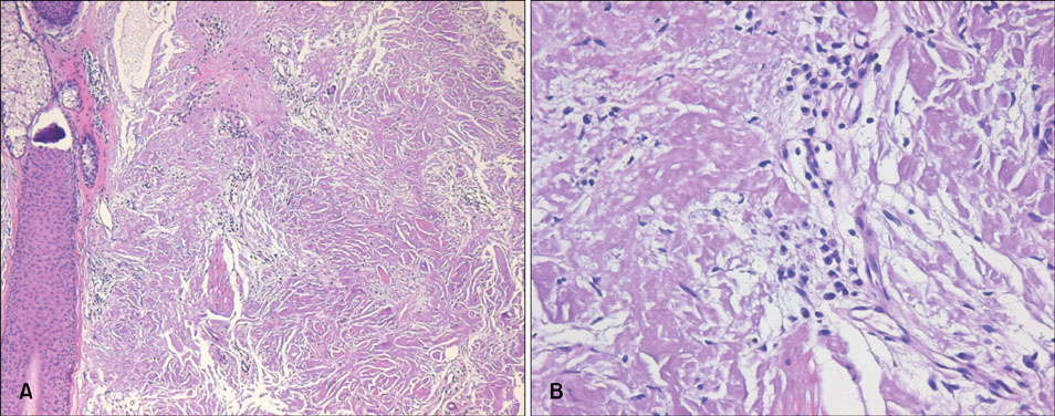
Fig. 2 (A) Deposition of acellular amorphous eosinophilic materials over the entire dermis (H&E, ×100). (B) Infiltration of numerous plasma cells within the deposits (H&E, ×400).

Fig. 3 Characteristic apple-green birefringence under polarized light (Congo red staining, ×200).
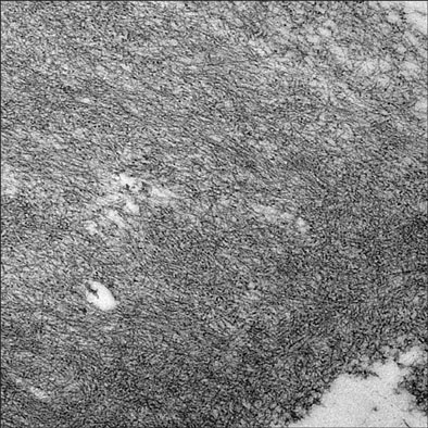
Fig. 4 Amyloid filaments are straight, long, and of uniform diameter (TEM, ×20,000).
Reference
-
1. Breathnach SM. Amyloid and amyloidosis. J Am Acad Dermatol. 1988. 18:1–16.
Article2. James WD, Berger TG, Elston DM. Andrew's diseases of the skin: clinical dermatology. 2006. 10th ed. Philadelphia: Saunders Elsevier;519–522.3. Elder DE, Elenitsas R, Johnson BL Jr, Murphy GF, Xu X. Lever's histopathology of the skin. 2009. 10th ed. Philadelphia: Lippincott Williams & Wilkins;870–876.4. Husby G, Sletten K, Blumenkrantz N, Danielsen L. Characterization of an amyloid fibril protein from localized amyloidosis of the skin as lambda immunoglobulin light chains of variable subgroup I (A lambda I). Clin Exp Immunol. 1981. 45:90–96.5. Kalajian AH, Waldman M, Knable AL. Nodular primary localized cutaneous amyloidosis after trauma: a case report and discussion of the rate of progression to systemic amyloidosis. J Am Acad Dermatol. 2007. 57:2 Suppl. S26–S29.6. Huilgol SC, Ramnarain N, Carrington P, Leigh IM, Black MM. Cytokeratins in primary cutaneous amyloidosis. Australas J Dermatol. 1998. 39:81–85.
Article7. Moon AO, Calamia KT, Walsh JS. Nodular amyloidosis: review and long-term follow-up of 16 cases. Arch Dermatol. 2003. 139:1157–1159.8. Masuda C, Mohri S, Nakajima H. Histopathological and immunohistochemical study of amyloidosis cutis nodularis atrophicans--comparison with systemic amyloidosis. Br J Dermatol. 1988. 119:33–43.
Article9. Rodermund OE. On amyloidosis cutis nodularis atrophicans (Gottron 1950). At the same time a contribution to the classification of amyloidoses. Arch Klin Exp Dermatol. 1967. 230:153–171.10. Dupre A, Bonafe JF, Pieraggi MT, Perrot H. Paracolloid of the skin. J Cutan Pathol. 1979. 6:304–309.
Article11. Kawashima Y, Matsubara T, Kinbara T, Hirone T, Kitamura K, Himi A, et al. Colloid degeneration of the skin--a case report. J Dermatol. 1977. 4:115–121.12. Kitajima Y, Seno J, Aoki S, Tada S, Yaoita H. Nodular primary cutaneous amyloidosis. Isolation and characterization of amyloid fibrils. Arch Dermatol. 1986. 122:1425–1430.
Article13. Symonds DA, Eichelberger MF, Sager GL. Calcifying amyloidoma of the breast. South Med J. 1995. 88:1169–1172.
Article14. Pasternak S, Wright BA, Walsh N. Soft tissue amyloidoma of the extremities: report of a case and review of the literature. Am J Dermatopathol. 2007. 29:152–155.15. Brownstein MH, Helwig EB. The cutaneous amyloidoses I Localized forms. Arch Dermatol. 1970. 102:8–19.
Article16. Woollons A, Black MM. Nodular localized primary cutaneous amyloidosis: a long-term follow-up study. Br J Dermatol. 2001. 145:105–109.
Article
- Full Text Links
-
- Actions
-
Cited
- CITED
-
- Close
- Share
-
- Similar articles
-
- Primary Localized Cutaneous Nodular Amyloidosis on Scalp Successfully Treated with Excision
- A Case of Familial Primary Localized Cutaneous Amyloidosis
- Primary Cutaneous Nodular Amyloidosis in Both Lip Angles
- A case of nodular amyloidosis
- Insulin-Derived Cutaneous Amyloidosis: A Possible Complication of Repeated Insulin Injections
