Management of Aneurysms of the Proximal (A1) Segment of the Anterior Cerebral Artery
- Affiliations
-
- 1Department of Neurosurgery, Busan-Ulsan Regional Cardiocerebrovascular Center, Medical Science Research Center, College of Medicine, Dong-A University, Busan, Korea. jthuh@dau.ac.kr
- 2Department of Diagnostic Radiology, Busan-Ulsan Regional Cardiocerebrovascular Center, Medical Science Research Center, College of Medicine, Dong-A University, Busan, Korea.
- KMID: 2172025
- DOI: http://doi.org/10.7461/jcen.2013.15.1.13
Abstract
OBJECTIVE
Aneurysms originating from the proximal segment (A1) of the anterior cerebral artery are rare; however, because of their small size, the risk of injury of perforating arteries, and the location of the aneurysm in the surgical field, they are challenging to treat. We report on 15 patients with A1 aneurysms and review surgical views according to the direction of aneurysms.
METHODS
Fifteen patients were diagnosed with A1 aneurysms and underwent surgical clipping or endovascular coiling at our institution between January 2006 and March 2012. We conducted a retrospective review of clinical and radiological features of all patients with A1 aneurysms.
RESULTS
Nine patients underwent surgical clipping, and six patients received endovascular coiling. Six patients (40%) had multiple aneurysms. A1 aneurysms ranged in size from 1.5 to 8.2 mm, with an average size of 3.26 mm. Most A1 aneurysms (73%) had a posterior direction. In the surgical view, A1 aneurysms projecting posteriorly were located behind the A1 trunk. The A1 aneurysm projecting posteroinferiorly was completely eclipsed by the parent artery. In A1 aneurysms with a posterosuperior or superior direction, finding and clipping the aneurysm neck was relatively easy. Thirteen patients (87%) had an excellent outcome, one had moderate disability, and one died.
CONCLUSION
A1 aneurysms have certain characteristics; small size, multiple aneurysms, and, usually, a posterior direction. A1 aneurysms with a posterosuperior or superior direction are relatively easy to assess, however, clipping of A1 aneurysms with a posterior or posteroinferior direction is more difficult. Endovascular coiling is an alternative therapeutic option when surgical clipping is expected to be difficult.
MeSH Terms
Figure
-
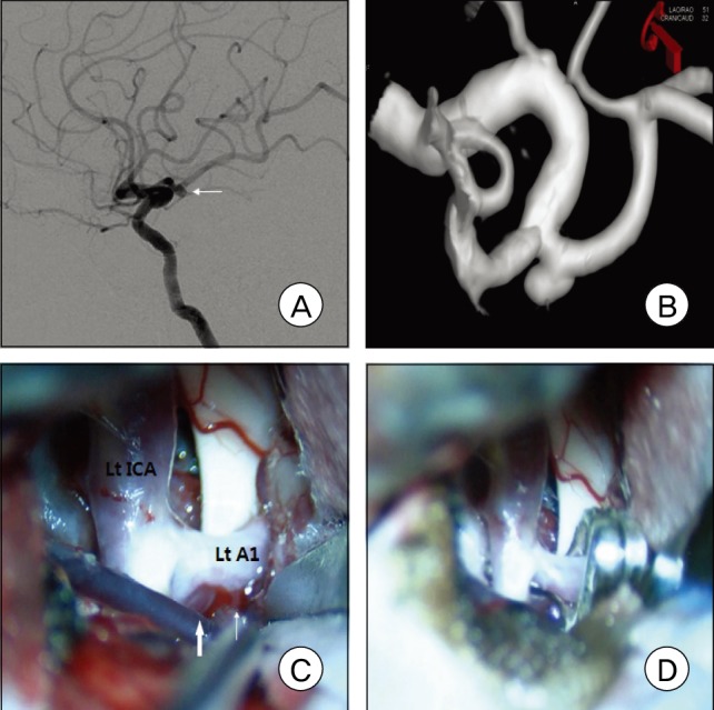
Fig. 1 A: Left carotid angiogram shows a left A1 aneurysm with a posterior projection. B: The surgical view from three-dimensional digital subtraction angiography was made for planning of aneurysm surgery C: Intraoperative photograph shows an A1 aneurysm (thick arrow) located behind the A1 trunk and a perforating artery (thin arrow). D: A fenestrated clip was applied to the aneurysm parallel to the parent artery.
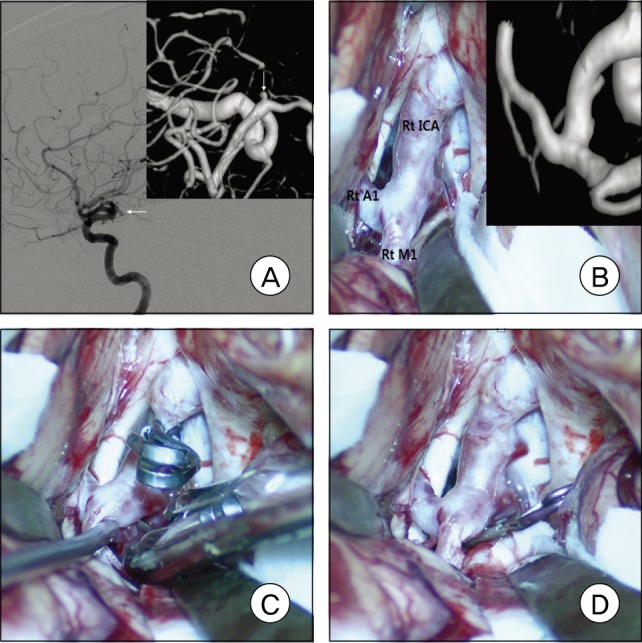
Fig. 2 A: Right carotid angiogram shows a right A1 aneurysm with a posteroinferior projection. B: Intraoperative photograph shows that the aneurysm was completely eclipsed by the parent artery, as shown in the surgical view from three-dimensional digital subtraction angiography. C and D: The aneurysm was found and clipped after mobilization of the internal carotid artery and middle cerebral artery.
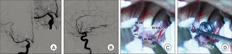
Fig. 3 A and B: Right carotid angiograms show a right A1 aneurysm with a posterosuperior projection. C and D: Intraoperative photographs show that finding and clipping the aneurysm neck was relatively easy.
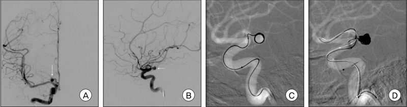
Fig. 4 A and B: Right carotid angiograms show a right A1 aneurysm with a posterior projection. C: The microcatheter kicked back out of the right A1 aneurysm during delivery of the first complex coil. D: Balloon-assisted coil embolization technique was used.
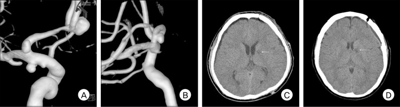
Fig. 5 A: Left carotid angiogram shows multiple A1 aneurysms (one of the anterior communicating artery, one of the left anterior choroidal artery, and one of the left A1). B: Left carotid angiogram shows a left A1 aneurysm. C and D: Postoperative computed tomography scans show a small area of low density in the genu portion of the internal capsule in each patient (A and B).
Reference
-
1. Chang HW, Youn SW, Jung C, Kang HS, Sohn CH, Kwon BJ, et al. Technical strategy in endovascular treatment of proximal anterior cerebral artery aneurysms. Acta Neurochir (Wien). 2011; 2. 153(2):279–285. PMID: 20872259.
Article2. Czepko R, Libionka W, Lopatka P. Characteristics and surgery of aneurysms of the proximal (A1) segment of the anterior cerebral artery. J Neurosurg Sci. 2005; 9. 49(3):85–95. PMID: 16288191.3. Dashti R, Hernesniemi J, Lehto H, Niemelä M, Lehecka M, Rinne J, et al. Microneurosurgical management of proximal anterior cerebral artery aneurysms. Surg Neurol. 2007; 10. 68(4):366–377. PMID: 17905060.
Article4. Dunker RO, Harris AB. Surgical anatomy of the proximal anterior cerebral artery. J Neurosurg. 1976; 3. 44(3):359–367. PMID: 1249614.
Article5. Gupta R, Horowitz MB, Gilman S. Neuroform stent-assisted coil embolization of a ruptured A1 segment anterior cerebral artery aneurysm. J Neuroimaging. 2006; 4. 16(2):117–119. PMID: 16629732.
Article6. Handa J, Nakasu Y, Matsuda M, Kyoshima K. Aneurysms of the proximal anterior cerebral artery. Surg Neurol. 1984; 11. 22(5):486–490. PMID: 6495158.
Article7. Hino A, Fujimoto M, Iwamoto Y, Oka H, Echigo T. Surgery of proximal anterior cerebral artery aneurysms. Acta Neurochir (Wien). 2002; 12. 144(12):1291–1296. discussion 1296. PMID: 12478340.8. Lee HY, Ahn JS, Suh DC, Lee DH. Z-shaped microcatheter tip shaping for embolization of aneurysms at the proximal A1 segment of the anterior cerebral artery: a technical note. Neurointervention. 2011; 8. 6(2):95–99. PMID: 22125756.
Article9. Lee JM, Joo SP, Kim TS, Go EJ, Choi HY, Seo BR. Surgical management of anterior cerebral artery aneurysms of the proximal (A1) segment. World Neurosurg. 2010; Oct-Nov. 74(4-5):478–482. PMID: 21492598.
Article10. Lubicz B, Bruneau M, Dewindt A, Lwfranc F, Baleriaux D, De Whitte O. Endovascular treatment of proximal anterior cerebral artery aneurysms. Neuroradiology. 2009; 2. 51(2):99–102. PMID: 18985332.
Article11. Perlmutter D, Rhoton AL Jr. Microsurgical anatomy of the anterior cerebral-anterior communicating-recurrent artery complex. J Neurosurg. 1976; 9. 45(3):259–272. PMID: 948013.
Article12. Raabe A, Beck J, Gerlach R, Zimmermann M, Seifert V. Near-infrared indocyanine green video angiography: a new method for intraoperative assessment of vascular flow. Neurosurgery. 2003; 1. 52(1):132–139. discussion 139. PMID: 12493110.
Article13. Rosner SS, Rhoton AL Jr, Ono M, Barry M. Microsurgical anatomy of the anterior perforating arteries. J Neurosurg. 1984; 9. 61(3):468–485. PMID: 6747683.
Article14. Suzuki M, Onuma T, Sakurai Y, Mizoi K, Ogawa A, Yoshimoto T. Aneurysms arising from the proximal (A1) segment of the anterior cerebral artery. A study of 38 cases. J Neurosurg. 1992; 3. 76(3):455–458. PMID: 1738027.
- Full Text Links
-
- Actions
-
Cited
- CITED
-
- Close
- Share
-
- Similar articles
-
- Aneurysms of the Proximal Anterior Cerebral Artery
- Aneurysms of Proximal(A1) Segment of Anterior Cerebral Artery
- Aneurysms of Proximal(A1) Segment of Anterior Cerebral Artery
- Ruptured Saccular Aneurysm Arising from Fenestrated Proximal Anterior Cerebral Artery : Case Report and Literature Review
- Microsurgical anatomy of the Anterior Cerebral-anterior Communicating Artery
