Ann Dermatol.
2016 Apr;28(2):247-248. 10.5021/ad.2016.28.2.247.
Cutaneous Metastasis of Giant Cell-Rich Osteosarcoma
- Affiliations
-
- 1Department of Dermatology, Seoul St. Mary's Hospital, College of Medicine, The Catholic University of Korea, Seoul, Korea. yymmpark6301@hotmail.com
- KMID: 2171386
- DOI: http://doi.org/10.5021/ad.2016.28.2.247
Abstract
- No abstract available.
MeSH Terms
Figure
-
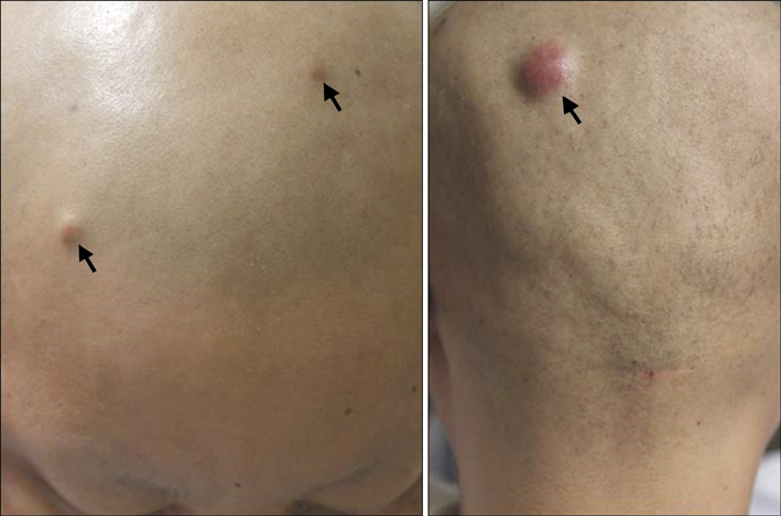
Fig. 1 Three 5 to 12-mm, flesh-toned to erythematous, firm nodules on the scalp (arrows).

Fig. 2 (A) Hypercellular stroma composed of mononuclear tumor cells and many multinucleated giant cells (H&E, ×40). (B) Mononuclear tumor cells with a variety of morphologies and multinucleated giant cells containing large pleomorphic nuclei (H&E, ×400). (C) Primary tumor of the right femur; numerous multinucleated giant cells and atypical tumor cells (H&E, ×400).
Reference
-
1. Park SG, Song JY, Song IG, Kim MS, Shin BS. Cutaneous extraskeletal osteosarcoma on the scar of a previous bone graft. Ann Dermatol. 2011; 23:Suppl 2. S160–S164.
Article2. Wang CS, Yin QH, Liao JS, Lou JH, Ding XY, Zhu YB. Giant cell-rich osteosarcoma in long bones: clinical, radiological and pathological features. Radiol Med. 2013; 118:1324–1334.
Article3. Huang J, Jiang Z, Zhang H. Clinicopathologic differential diagnosis of giant cell-rich osteosarcoma and giant cell tumor of bone. Zhonghua Bing Li Xue Za Zhi. 2014; 43:379–382.4. Mariano FV, Corrêa MB, da Costa MV, de Almeida OP, Lopes MA. Labial mucosa metastasis of fibule giant cell-rich osteosarcoma: an unusual presentation. Quintessence Int. 2013; 44:783–791.5. Oliveira AM, Dei Tos AP, Fletcher CD, Nascimento AG. Primary giant cell tumor of soft tissues: a study of 22 cases. Am J Surg Pathol. 2000; 24:248–256.
- Full Text Links
-
- Actions
-
Cited
- CITED
-
- Close
- Share
-
- Similar articles
-
- Giant Cell-Rich Osteosarcoma: A Tumor Simulating Borderline Lesion
- Denosumab-Treated Giant Cell Tumor of the Bone Mimicking Low-Grade Central Osteosarcoma
- Giant Cell Tumor with an Unusual Cartilage Matrix: A Case Report
- Malignant Transformation of Benign Giant Cell Tumor
- Osteocalcin expression in primary bone tumors: in situ hybridization and immunohistochemical study
