A Case of Cervical Retrotracheal Metastatic Papillary Thyroid Carcinoma Diagnosed by Endobronchial Ultrasonography with Transbronchial Needle Aspiration
- Affiliations
-
- 1Department of Otorhinolaryngology-Head and Neck Surgery, Kosin University Gospel Hospital, Busan, Korea. kdlee59@gmail.com
- 2Department of Internal Medicine, Kosin University Gospel Hospital, Busan, Korea.
- KMID: 2171243
- DOI: http://doi.org/10.11106/ijt.2015.8.2.235
Abstract
- A 61-year-old woman who underwent total thyroidectomy for papillary thyroid carcinoma (PTC) five years previously referred for a cervical retrotracheal mass. The mass had intense fluorodeoxyglucose (FDG) uptake on positron emission tomography-computed tomography (PET-CT), and was thus thought to be malignant. Transcutaneous ultrasonography with fine needle aspiration (FNA) was not feasible, so we tried endobronchial ultrasonography (EBUS) with transbronchial needle aspiration (TBNA) to obtain a cytology specimen. After surgery, the mass was confirmed to be a metastatic lymph node from the previous PTC, confirming the TBNA results. Although the utility of EBUS-TBNA for evaluating mediastinal metastasis has been reported in a number of studies, few reports have addressed its utility in the cervical region. Here we report this unusual case of metastatic lymph node of PTC that recurred in the cervical retrotracheal area. It was found to exhibit esophageal muscular invasion, and was accurately diagnosed on EBUS-TBNA.
MeSH Terms
Figure
-
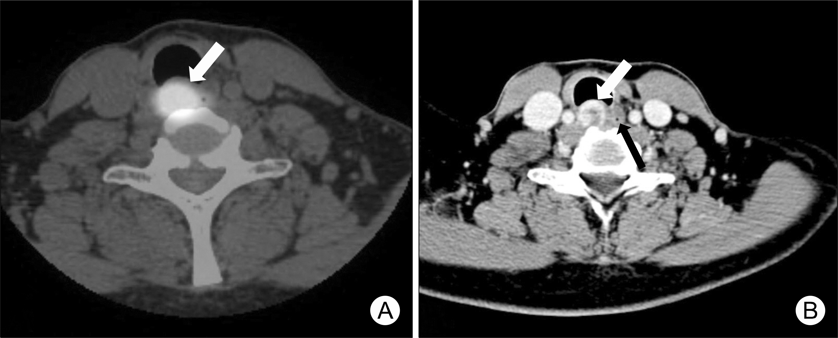
Fig. 1. PET-CT and CT scan of retrotracheal mass. (A) PET-CT scan showed fluoro-deoxyglucose uptake in the retrotracheal mass (arrow). (B) CT scan showed well-cir-cumscribed enhancing mass (white arrow) with central low density adjacent to the eso-phagus (black arrow). The mass was compressing the trachea anteriorly.
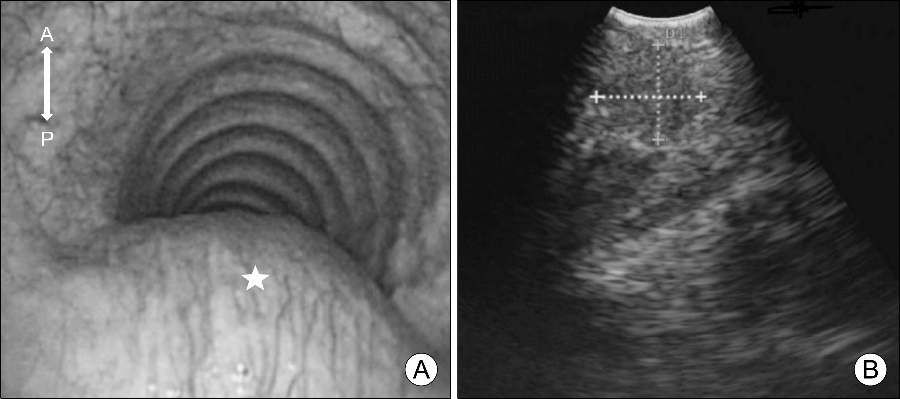
Fig. 2. EBUS images. (A) A large mass (star) was visua-lized protruding into the pos-terior trachea wall in endob-ronchial view (A: anterior, P: posterior). (B) In EBUS image, 1×1 cm retrotracheal mass with a hypoechogenic ap-pearance was seen.
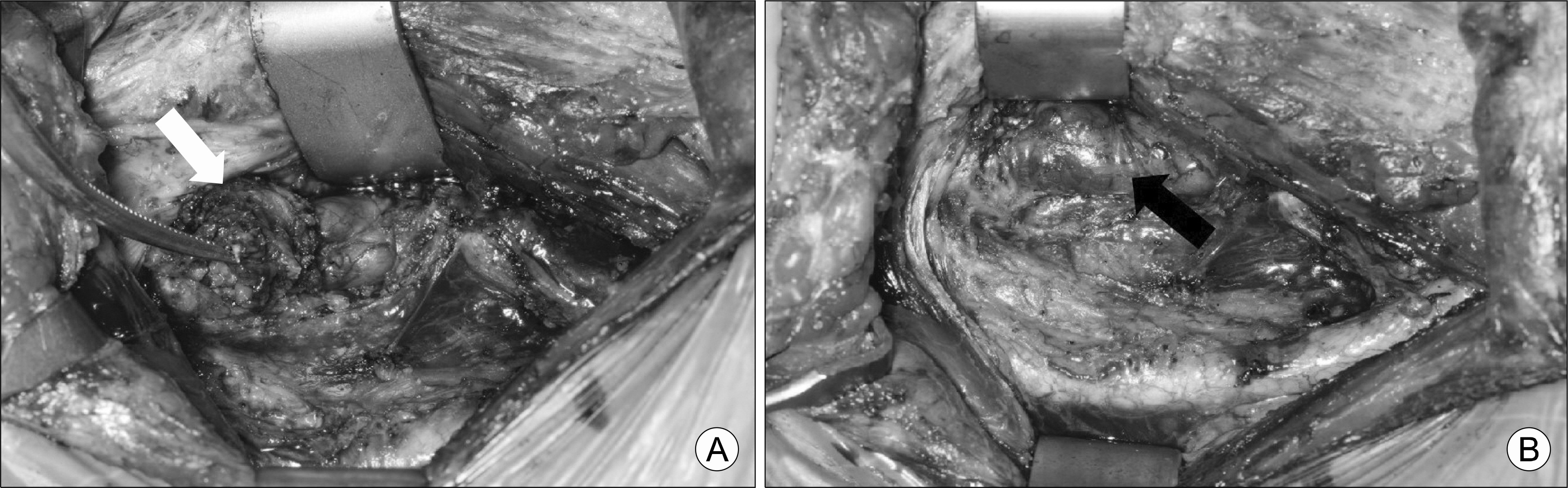
Fig. 3. Intraoperative view. (A) The mass (white arrow) was found to be grossly adherent to the trachea anteriorly and to the esophagus laterally. (B) The inner esophageal mucosa (black arrow) was intact after the mass was removed.
Reference
-
References
1. McCaffrey JC. Aerodigestive tract invasion by well-differentiated thyroid carcinoma: diagnosis, management, prognosis, and biology. Laryngoscope. 2006; 116(1):1–11.
Article2. Choi JS, Kim J, Kwak JY, Kim MJ, Chang HS, Kim EK. Preoperative staging of papillary thyroid carcinoma: comparison of ultrasound imaging and CT. AJR Am J Roentgenol. 2009; 193(3):871–8.
Article3. Yasufuku K, Chiyo M, Sekine Y, Chhajed PN, Shibuya K, Iizasa T, et al. Realtime endobronchial ultrasound-guided transbronchial needle aspiration of mediastinal and hilar lymph nodes. Chest. 2004; 126(1):122–8.
Article4. Yasufuku K, Chiyo M, Koh E, Moriya Y, Iyoda A, Sekine Y, et al. Endobronchial ultrasound guided transbronchial needle aspiration for staging of lung cancer. Lung Cancer. 2005; 50(3):347–54.
Article5. Yasufuku K, Nakajima T, Fujiwara T, Chiyo M, Iyoda A, Yoshida S, et al. Role of endobronchial ultrasound-guided transbronchial needle aspiration in the management of lung cancer. Gen Thorac Cardiovasc Surg. 2008; 56(6):268–76.
Article6. Machens A, Holzhausen HJ, Dralle H. Skip metastases in thyroid cancer leaping the central lymph node compartment. Arch Surg. 2004; 139(1):43–5.
Article7. Sivanandan R, Soo KC. Pattern of cervical lymph node metastases from papillary carcinoma of the thyroid. Br J Surg. 2001; 88(9):1241–4.
Article8. Kupferman ME, Patterson M, Mandel SJ, LiVolsi V, Weber RS. Patterns of lateral neck metastasis in papillary thyroid carcinoma. Arch Otolaryngol Head Neck Surg. 2004; 130(7):857–60.
Article9. Lupoli GA, Fonderico F, Colarusso S, Panico A, Cavallo A, Di Micco L, et al. Current management of differentiated thyroid carcinoma. Med Sci Monit. 2005; 11(12):RA368–73.10. McCaffrey JC. Evaluation and treatment of aerodigestive tract invasion by well-differentiated thyroid carcinoma. Cancer Control. 2000; 7(3):246–52.
Article11. Hurter T, Hanrath P. Endobronchial sonography: feasibility and preliminary results. Thorax. 1992; 47(7):565–7.
Article12. Yasufuku K, Nakajima T, Motoori K, Sekine Y, Shibuya K, Hiroshima K, et al. Comparison of endobronchial ultrasound, positron emission tomography, and CT for lymph node staging of lung cancer. Chest. 2006; 130(3):710–8.
Article13. Herth FJ, Ernst A, Eberhardt R, Vilmann P, Dienemann H, Krasnik M. Endobronchial ultrasound-guided transbronchial needle aspiration of lymph nodes in the radiologically normal mediastinum. Eur Respir J. 2006; 28(5):910–4.
Article14. Herth F, Becker HD, Ernst A. Conventional vs endobronchial ultrasound-guided transbronchial needle aspiration: a randomized trial. Chest. 2004; 125(1):322–5.15. Herth FJ, Becker HD, Ernst A. Ultrasound-guided transbronchial needle aspiration: an experience in 242 patients. Chest. 2003; 123(2):604–7.16. Machens A, Hinze R, Lautenschlager C, Thomusch O, Dralle H. Thyroid carcinoma invading the cervicovisceral axis: routes of invasion and clinical implications. Surgery. 2001; 129(1):23–8.
Article17. McCaffrey TV, Bergstralh EJ, Hay ID. Locally invasive papillary thyroid carcinoma: 1940–1990. Head Neck. 1994; 16(2):165–72.
Article18. Gillenwater AM, Goepfert H. Surgical management of laryngotracheal and esophageal involvement by locally advanced thyroid cancer. Semin Surg Oncol. 1999; 16(1):19–29.
Article
- Full Text Links
-
- Actions
-
Cited
- CITED
-
- Close
- Share
-
- Similar articles
-
- Oxyphilic Papillary Carcinoma of the Thyroid in Fine Needle Aspiration
- Multiple Cervical Schwannomas Mimicking Metastatic Lymph Nodes from Papillary Thyroid Cancer
- A Case of Mediastinal Ectopic Thyroid Tissue Diagnosed by Endobronchial Ultrasound Guided Transbronchial Needle Aspiration
- Concurrent Papillary and Medullary Carcinoma of the Thyroid Gland
- Fine needle aspiration cytology of mixed squamous cell carcinoma and papillary carcinoma in thyroid: a case report
