J Bone Metab.
2014 Nov;21(4):243-247. 10.11005/jbm.2014.21.4.243.
Assessing Bone Quality in Terms of Bone Mineral Density, Buckling Ratio and Critical Fracture Load
- Affiliations
-
- 1Department of Biomedical Engineering, National University of Singapore, Singapore.
- 2Department of Medical Biotechnology, Dongguk University, Seoul, Korea. tlee@dongguk.edu
- KMID: 2170051
- DOI: http://doi.org/10.11005/jbm.2014.21.4.243
Abstract
- BACKGROUND
Bone mineral density (BMD) is used as a sole parameter in the diagnosis of osteoporosis. Due to the ease of acquirement of BMD, clinical diagnosis still involves its usage although the limitations of BMD are quite well-established. Therefore, this preliminary study hoped to reduce the errors introduced by BMD alone by incorporating geometric and structural predictors simultaneously to observe if strength was implicitly dependent on the geometry and BMD. Hence, we illustrated the triadic relationship between BMD, buckling ratio (BR) and critical fracture load (Fcr).
METHODS
The geometric predictor was the BR as it involves both the changes in the periosteum and the cortical thickness. Also, structural changes were monitored by finite element (FE) analysis-predicted Fcr. These BR and Fcr measurements were plotted with their respective femoral neck BMD values in elderly female patients (n=6) in a 3-year follow-up study, treated with ibandronate.
RESULTS
In all the three-dimensional plots (baseline, mid and final year), high Fcr values were found at regions containing high BMD and low BR values. Quantitatively, this was also proven where an averaged highest Fcr across the three years had a relatively higher BMD (46%) and lower BR (19%) than that of the averaged lowest Fcr. The dependence of FE predicted strength on both the geometry and bone density was illustrated.
CONCLUSIONS
We conclude that use of triadic relationships for the evaluation of osteoporosis and hip fractures with the combination of strength, radiology-derived BR and bone density will lay the foundation for more accurate predictions in the future.
MeSH Terms
Figure
-

Fig. 1 Obtainment of outer radius and cortical thickness from profile rays drawn on narrowest neck slice. After extraction of femoral neck region, profile rays were drawn at 30° intervals, measured from the centroid of the femoral neck slice. A sample profile ray at 0° is shown where respective regions of cortical bone, trabecular bone and soft tissues are identified.
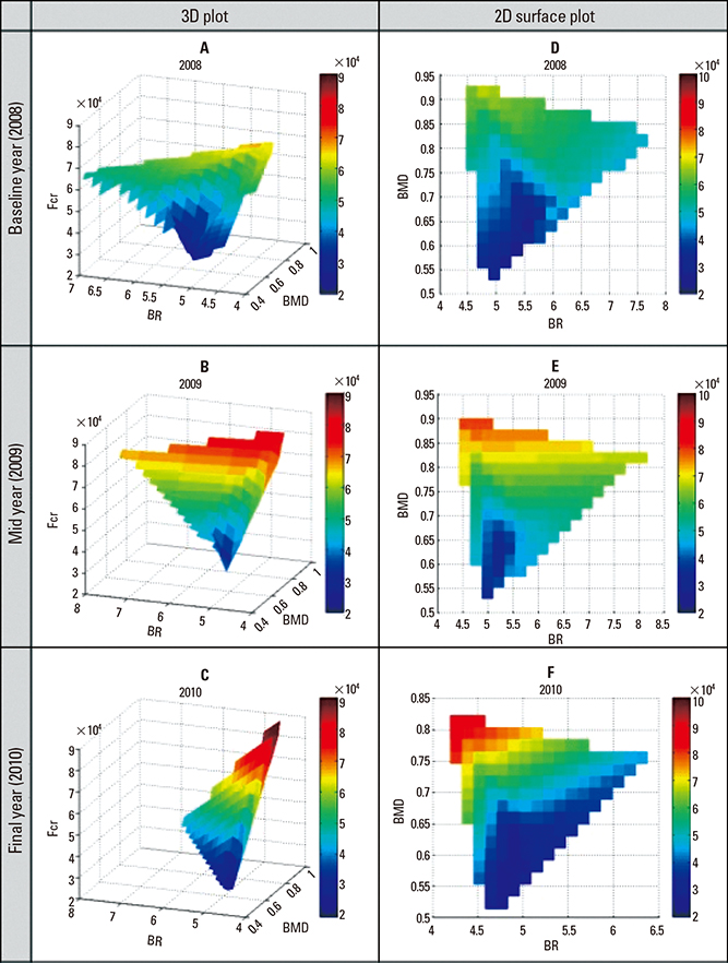
Fig. 2 Yearly three-dimensional surface plots of bone mineral density (BMD), buckling ratio and fracture load (Fcr). High Fcr values were found at regions containing high BMD and low BR values.
Reference
-
1. Melton LJ 3rd. Hip fractures: a worldwide problem today and tomorrow. Bone. 1993; 14:Suppl 1. S1–S8.
Article2. Kanis JA, Melton LJ 3rd, Christiansen C, et al. The diagnosis of osteoporosis. J Bone Miner Res. 1994; 9:1137–1141.
Article3. Geusens P, van Geel T, van den Bergh J. Can Hip Fracture Prediction in Women be Estimated beyond Bone Mineral Density Measurement Alone? Ther Adv Musculoskelet Dis. 2010; 2:63–77.
Article4. Schuit SC, van der Klift M, Weel AE, et al. Fracture incidence and association with bone mineral density in elderly men and women: the Rotterdam Study. Bone. 2004; 34:195–202.
Article5. Anitha D, Kim KJ, Lim SK, et al. Implications of local osteoporosis on the efficacy of anti-resorptive drug treatment: a 3-year follow-up finite element study in risedronate-treated women. Osteoporos Int. 2013; 24:3043–3051.
Article6. Lotz JC, Cheal EJ, Hayes WC. Fracture prediction for the proximal femur using finite element models: part I--Linear analysis. J Biomech Eng. 1991; 113:353–360.
Article7. Bessho M, Ohnishi I, Matsuyama J, et al. Prediction of strength and strain of the proximal femur by a CT-based finite element method. J Biomech. 2007; 40:1745–1753.
Article8. Schileo E, Taddei F, Malandrino A, et al. Subject-specific finite element models can accurately predict strain levels in long bones. J Biomech. 2007; 40:2982–2989.
Article9. Cristofolini L, Schileo E, Juszczyk M, et al. Mechanical testing of bones: the positive synergy of finite-element models and in vitro experiments. Philos Trans A Math Phys Eng Sci. 2010; 368:2725–2763.
Article10. Beck TJ, Oreskovic TL, Stone KL, et al. Structural adaptation to changing skeletal load in the progression toward hip fragility: the study of osteoporotic fractures. J Bone Miner Res. 2001; 16:1108–1119.
Article11. Bouxsein ML. Determinants of skeletal fragility. Best Pract Res Clin Rheumatol. 2005; 19:897–911.
Article12. Carpenter RD, Sigurdsson S, Zhao S, et al. Effects of age and sex on the strength and cortical thickness of the femoral neck. Bone. 2011; 48:741–747.
Article13. Keyak JH. Improved prediction of proximal femoral fracture load using nonlinear finite element models. Med Eng Phys. 2001; 23:165–173.
Article14. Johnell O, Kanis JA, Oden A, et al. Predictive value of BMD for hip and other fractures. J Bone Miner Res. 2005; 20:1185–1194.
Article15. Cawthon PM, Ewing SK, Mackey DC, et al. Change in hip bone mineral density and risk of subsequent fractures in older men. J Bone Miner Res. 2012; 27:2179–2188.
Article16. Parfitt AM. Skeletal heterogeneity and the purpose of bone remodeling: implications for the understanding of osteoporosis. In : Marcus R, Feldman D, Kelsey JL, editors. Osteoporosis. 2nd ed. San Diego, CA: Academic Press;2001. p. 433–447.
- Full Text Links
-
- Actions
-
Cited
- CITED
-
- Close
- Share
-
- Similar articles
-
- Osteoporosis
- The Usefulness of the Dual Energy X-ray Absorptiometry(DEXA) for the Bone Density of the Tricortical Iliac Graft in the Spinal Interbody Fusion
- Diffrences of Bone Mineral Density between Osteoporotic Group with or without Compression Fracture of the Spine
- Osteoporotic Ankle Fracture
- Diabetes and bone
