Imaging Sci Dent.
2013 Sep;43(3):185-190. 10.5624/isd.2013.43.3.185.
Effect of digital noise reduction on the accuracy of endodontic file length determination
- Affiliations
-
- 1Department of Oral and Maxillofacial Radiology, School of Dentistry, Isfahan University of Medical Sciences, Isfahan, Iran. nastaranfarhadi@yahoo.com
- 2Department of Endodontics, School of Dentistry, Isfahan University of Medical Sciences, Isfahan, Iran.
- KMID: 2167456
- DOI: http://doi.org/10.5624/isd.2013.43.3.185
Abstract
- PURPOSE
The aim of the present study was to evaluate the measurement accuracy of endodontic file length on periapical digital radiography after application of noise reduction digital enhancement.
MATERIALS AND METHODS
Thirty-five human single-rooted permanent teeth with canals measuring 20-24 mm in length were selected. ISO #08 endodontic files were placed in the root canals of the teeth. The file lengths were measured with a digital caliper as the standard value. Standard periapical digital images were obtained using the Digora digital radiographic system and a dental X-ray unit. In order to produce the enhanced images, the noise reduction option was applied. Two blinded radiologists measured the file lengths on the original and enhanced images. The measurements were compared by repeated measures ANOVA and the Bonferroni test (alpha=0.05).
RESULTS
Both the original and enhanced digital images provided significantly longer measurements compared with the standard value (P<0.05). There were no significant differences between the measurement accuracy of the original and enhanced images (P>0.05).
CONCLUSION
Noise reduction digital enhancement did not influence the measurement accuracy of the length of the thin endodontic files on the digital periapical radiographs despite the fact that noise reduction could result in the elimination of fine details of the images.
MeSH Terms
Figure
-

Fig. 1 A. Insertion of an endodontic file into the canal. B. File length measurement (standard value). C. Placement of tooth into cadaver socket. D. Scanora software. E. Original image. F. enhanced image.
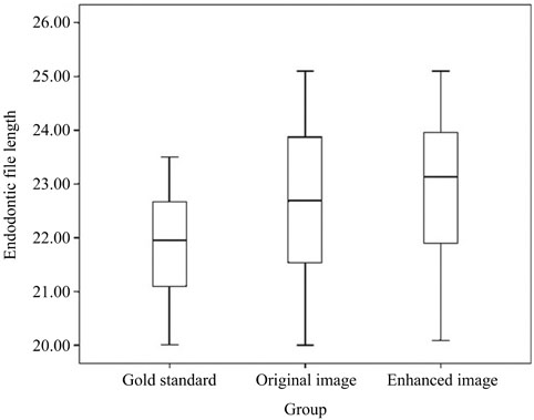
Fig. 2 Box plot of endodontic file length in the standard value and on original and enhanced digital radiographs.
Reference
-
1. Vandenberghe B, Jacobs R, Bosmans H. Modern dental imaging: a review of the current technology and clinical applications in dental practice. Eur Radiol. 2010; 20:2637–2655.
Article2. Gormez O, Yilmaz HH. Image post-processing in dental practice. Eur J Dent. 2009; 3:343–347.
Article3. Parks ET, Williamson GF. Digital radiography: an overview. J Contemp Dent Pract. 2002; 3:23–39.
Article4. Almenar Garcia A, Forner Navarro L, Ubet Castello V, Minana Laliga R. Evaluation of a digital radiography to estimate working length. J Endod. 1997; 23:363–365.5. Kositbowornchai S, Nuansakul R, Sikram S, Sinahawattana S, Saengmontri S. Root fracture detection: a comparison of direct digital radiography with conventional radiography. Dentomaxillofac Radiol. 2001; 30:106–109.
Article6. Kamburoğlu K, Barenboim SF, Kaffe I. Comparison of conventional film with different digital and digitally filtered images in the detection of simulated internal resorption cavities - an ex vivo study in human cadaver jaws. Oral Surg Oral Med Oral Pathol Oral Radiol Endod. 2008; 105:790–797.7. Borg E, Gröndahl K, Persson LG, Gröndahl HG. Marginal bone level around implants assessed in digital and film radiographs: in vivo study in the dog. Clin Implant Dent Relat Res. 2000; 2:10–17.
Article8. Syriopoulos K, Sanderink GC, Velders XL, van der Stelt PF. Radiographic detection of approximal caries: a comparison of dental films and digital imaging systems. Dentomaxillofac Radiol. 2000; 29:312–318.
Article9. Akdeniz B, Soğur E. An ex vivo comparison of conventional and digital radiography for perceived image quality of root fillings. Int Endod J. 2005; 38:397–401.
Article10. Naoum HJ, Chandler NP, Love RM. Conventional versus storage phosphor-plate digital images to visualize the root canal system contrasted with a radiopaque medium. J Endod. 2003; 29:349–352.
Article11. Kavadella A, Karayiannis A, Nicopoulou-Karayianni K. Detectability of experimental peri-implant cancellous bone lesions using conventional and direct digital radiography. Aust Dent J. 2006; 51:180–186.
Article12. Mohtavipour ST, Dalili Z, Azar NG. Direct digital radiography versus conventional radiography for estimation of canal length in curved canals. Imaging Sci Dent. 2011; 41:7–10.
Article13. Li G. Comparative investigation of subjective image quality of digital intraoral radiographs processed with 3 image-processing algorithms. Oral Surg Oral Med Oral Pathol Oral Radiol Endod. 2004; 97:762–767.
Article14. van der Stelt PF. Filmless imaging: the uses of digital radiography in dental practice. J Am Dent Assoc. 2005; 136:1379–1387.15. van der Stelt PF. Better imaging: the advantages of digital radiography. J Am Dent Assoc. 2008; 139:Suppl. 7S–13S.16. Brüllmann DD, Röhrig B, Sulayman SL, Schulze R. Length of endodontic files measured in digital radiographs with and without noise-suppression filters: an ex-vivo study. Dentomaxillofac Radiol. 2011; 40:170–176.17. Castro V, Katz J, Hardman P, Glaros A, Spencer P. In vitro comparison of conventional film and direct digital imaging in the detection of approximal caries. Dentomaxillofac Radiol. 2007; 36:138–142.18. Eickholz P, Riess T, Lenhard M, Hassfeld S, Staehle HJ. Digital radiography of interproximal bone loss; validity of different filters. J Clin Periodontol. 1999; 26:294–300.
Article19. Kal BI, Baksi BG, Dündar N, Sen BH. Effect of various digital processing algorithms on the measurement accuracy of endodontic file length. Oral Surg Oral Med Oral Pathol Oral Radiol Endod. 2007; 103:280–284.
Article20. Koob A, Sanden E, Hassfeld S, Staehle HJ, Eickholz P. Effect of digital filtering on the measurement of the depth of proximal caries under different exposure conditions. Am J Dent. 2004; 17:388–393.21. Sund T, Møystad A. Sliding window adaptive histogram equalization of intraoral radiographs: effect on image quality. Dentomaxillofac Radiol. 2006; 35:133–138.
Article22. Møystad A, Svanaes DB, Risnes S, Larheim TA, Gröndahl HG. Detection of approximal caries with a storage phosphor system. A comparison of enhanced digital images with dental X-ray film. Dentomaxillofac Radiol. 1996; 25:202–206.
Article23. Raitz R, Assunção Junior JN, Fenyo-Pereira M, Correa L, de Lima LP. Assessment of using digital manipulation tools for diagnosing mandibular radiolucent lesions. Dentomaxillofac Radiol. 2012; 41:203–210.
Article24. Shrout MK, Russell C, Potter B, Powell B, Hildebolt C. Digital enhancement of radiographs: can it improve caries diagnosis? J Am Dent Assoc. 1996; 127:469–473.
Article25. de Molon RS, Morais-Camillo JA, Sakakura CE, Ferreira MG, Loffredo LC, Scaf G. Measurements of simulated periodontal bone defects in inverted digital image and film-based radiograph: an in vitro study. Imaging Sci Dent. 2012; 42:243–247.
Article26. Davies ER. Edge location shifts produced by median filters: theoretical bounds and experimental results. Signal Processing. 1989; 16:83–96.
Article27. Davies ER. Median and mean filters produce similar shifts on curved boundaries. Electron Lett. 1991; 27:826–828.
Article28. Davies ER. Formulation of an accurate discrete theory of median shifts. Signal Processing. 2003; 83:531–544.
Article29. Haak R, Wicht MJ. Grey-scale reversed radiographic display in the detection of approximal caries. J Dent. 2005; 33:65–71.
Article30. Xu Y, Lai EM. Restoration of images contaminated by mixed Gaussian and impulse noise using a recursive minimum-maximum method. IEE Proc Vis Image Signal Process. 1998; 145:264–270.
Article31. Nair MK, Nair UP. Digital and advanced imaging in endodontics: a review. J Endod. 2007; 33:1–6.
Article32. Brüllmann D, Witzel V, Willershausen B, d'Hoedt B. Effect of digital noise filters on diagnostic radiographs for the diagnosis of experimental root fractures. Int J Comput Dent. 2008; 11:107–114.33. Yalcinkaya S, Künzel A, Willers R, Thoms M, Becker J. Subjective image quality of digitally filtered radiographs acquired by the Dürr Vistascan system compared with conventional radiographs. Oral Surg Oral Med Oral Pathol Oral Radiol Endod. 2006; 101:643–651.
Article34. Näslund EB, Møystad A, Larheim TA, Øgaard B, Kruger M. Cephalometric analysis with digital storage phosphor images: extreme low-exposure images with and without postprocessing noise reduction. Am J Orthod Dentofacial Orthop. 2003; 124:190–197.
Article35. Mehdizadeh M, Khademi AA, Nasr N. Canal length measurement by digital radiography and conventional parallel radiography. Res J Biol Sci. 2010; 5:400–403.
Article36. Williams CB, Joyce AP, Roberts S. A comparison between in vivo radiographic working length determination and measurement after extraction. J Endod. 2006; 32:624–627.
Article37. Schmitd LB, Lima Tde C, Chinellato LE, Bramante CM, Garcia RB, de Moraes IG, et al. Comparison of radiographic measurements obtained with conventional and indirect digital imaging during endontic treatment. J Appl Oral Sci. 2008; 16:167–170.38. Brito-Júnior M, Santos LA, Baleeiro EN, Pêgo MM, Eleutério NB, Camilo CC. Linear measurements to determine working length of curved canals with fine files: conventional versus digital radiography. J Oral Sci. 2009; 51:559–564.
Article39. Macdonald R. Digital imaging for dentists. Aust Dent J. 2001; 46:301–305.
Article
- Full Text Links
-
- Actions
-
Cited
- CITED
-
- Close
- Share
-
- Similar articles
-
- Study of endodontic working length of Korean posterior teeth
- The accuracy of the radiographic method in root canal length measurement
- Comparison of three digital radiographic imaging systems for the visibility of endodontic files
- Direct digital radiography versus conventional radiography for estimation of canal length in curved canals
- Comparison of vibration characteristics of file systems for root canal shaping according to file length
