J Korean Acad Conserv Dent.
2010 Nov;35(6):429-435. 10.5395/JKACD.2010.35.6.429.
Study of endodontic working length of Korean posterior teeth
- Affiliations
-
- 1Department of Conservative Dentistry, Kyung Hee University School of Dentistry, Seoul, Korea. shpark94@khu.ac.kr
- 2Medical IS Team, Kyung Hee Management Company, Seoul, Korea.
- 3Department of Dental Hygiene, Wonkwang Health Science University, Iksan, Korea.
- 4Department of Conservative Dentistry, Kyung Hee University School of Dentistry, Seoul, Korea.
- KMID: 2176464
- DOI: http://doi.org/10.5395/JKACD.2010.35.6.429
Abstract
OBJECTIVES
The aim of this study was to investigate average working lengths of Korean posterior teeth and evaluate validity of endodontic file length.
MATERIALS AND METHODS
The endodontic working length of the posterior teeth of 670 Korean patients were measured than each mean value and standard deviation were investigated than the frequency deviation and standard deviation per each length were calculated.
RESULTS
Among the canals of premolar, 66.5% of canal length was marked under 20 mm by endodontic working length and 95.4% could be measured under 22 mm and Among the canals of molars, 95.5% of canal length was marked under 20 mm endodontic working length.
CONCLUSIONS
With the result of measurement of endodontic working length of premolars of Korean, it suggested that 23 mm endodontic file is more proper than the 21 mm and 25 mm file on the market.
Keyword
Figure
-
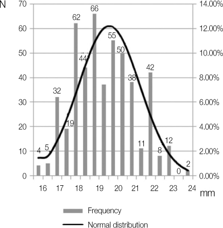
Figure 1 The number of canals classified by length in premolar. Frequency distribution and normal distribution.

Figure 2 The number of canals classified by length in molar. Frequency distribution and normal distribution.
Reference
-
1. Kim S. Modern endodontic practice: instruments and techniques. Dent Clin North Am. 2004. 48(1):1–9.
Article2. Barbakow F. The LightSpeed system. Dent Clin North Am. 2004. 48(1):113–135.
Article3. Clauder T, Baumann MA. ProTaper NT system. Dent Clin North Am. 2004. 48(1):87–111.
Article4. Hsu YY, Kim S. The ProFile system. Dent Clin North Am. 2004. 48(1):69–85.
Article5. Mounce RE. The K3 rotary nickel-titanium file system. Dent Clin North Am. 2004. 48(1):137–157.
Article6. Takatsuka S, Yoshida K, Ueki K, Marukawa K, Nakagawa K, Yamamoto E. Disc and condyle translation in patients with temporomandibular disorder. Oral Surg Oral Med Oral Pathol Oral Radiol Endod. 2005. 99(5):614–621.
Article7. Nunez SC, Garcez AS, Suzuki SS, Ribeiro MS. Management of mouth opening in patients with temporomandibular disorders through low-level laser therapy and transcutaneous electrical neural stimulation. Photomed Laser Surg. 2006. 24(1):45–49.
Article8. Kuttler Y. Microscopic investigation of root apexes. J Am Dent Assoc. 1955. 50(5):544–552.
Article9. Eliasson S, Lavstedt S, Ljungheimer C. Radiographic study of alveolar bone height related to tooth and root length. Community Dent Oral Epidemiol. 1986. 14(3):169–171.
Article10. Forsberg J. Radiographic reproduction of endodontic "working length" comparing the paralleling and the bisecting-angle techniques. Oral Surg Oral Med Oral Pathol. 1987. 64(3):353–360.
Article11. Burch JG, Hulen S. The relationship of the apical foramen to the anatomic apex of the tooth root. Oral Surg Oral Med Oral Pathol. 1972. 34(2):262–268.
Article12. Dummer PM, McGinn JH, Rees DG. The position and topography of the apical canal constriction and apical foramen. Int Endod J. 1984. 17(4):192–198.
Article13. Kim E, Lee SJ. Electronic apex locator. Dent Clin North Am. 2004. 48(1):35–54.
Article14. Vajrabhaya L, Tepmongkol P. Accuracy of apex locator. Endod Dent Traumatol. 1997. 13(4):180–182.
Article15. Czerw RJ, Fulkerson MS, Donnelly JC, Walmann JO. In vitro evaluation of the accuracy of several electronic apex locators. J Endod. 1995. 21(11):572–575.
Article16. Ibarrola JL, Chapman BL, Howard JH, Knowles KI, Ludlow MO. Effect of preflaring on Root ZX apex locators. J Endod. 1999. 25(9):625–626.
Article17. Shabahang S, Goon WW, Gluskin AH. An in vivo evaluation of Root ZX electronic apex locator. J Endod. 1996. 22(11):616–618.
Article18. Cho YL, Son WH, Hwnag HG. An accuracy of the several electronic apex locators on the mesial root canal of the mandibular molar. J Korean Acad Conserv Dent. 2005. 30(6):477–485.
Article19. Kim E, Fallahrastegar A, Hur YY, Jung IY, Kim S, Lee SJ. Difference in root canal length between Asians and Caucasians. Int Endod J. 2005. 38(3):149–151.
Article20. Perzigian AJ. Allometric analysis of dental variation in a human population. Am J Phys Anthropol. 1981. 54(3):341–345.
Article21. Cho JI, Jin MW, Kim YK, Kim SK. Change of working length in curved canals by various instrumentation techniques. J Korean Acad Conserv Dent. 2006. 31(1):30–35.
Article22. Gulabivala K, Aung TH, Alavi A, Ng YL. Root and canal morphology of Burmese mandibular molars. Int Endod J. 2001. 34(5):359–370.
Article23. Ng YL, Aung TH, Alavi A, Gulabivala K. Root and canal morphology of Burmese maxillary molars. Int Endod J. 2001. 34(8):620–630.
Article24. Ahmed HA, Abu-bakr NH, Yahia NA, Ibrahim YE. Root and canal morphology of permanent mandibular molars in a Sudanese population. Int Endod J. 2007. 40(10):766–771.
Article25. Pattanshetti N, Gaidhane M, Al Kandari AM. Root and canal morphology of the mesiobuccal and distal roots of permanent first molars in a Kuwait population-a clinical study. Int Endod J. 2008. 41(9):755–762.
Article26. Peiris HR, Pitakotuwage TN, Takahashi M, Sasaki K, Kanazawa E. Root canal morphology of mandibular permanent molars at different ages. Int Endod J. 2008. 41(10):828–835.
Article27. Park SH, Noh BH, Hwang HG. A Comparison of the length between mesio-buccal and mesio-lingual canals of the mandibular molar. J Korean Acad Conserv Dent. 2004. 29(6):541–547.
Article
- Full Text Links
-
- Actions
-
Cited
- CITED
-
- Close
- Share
-
- Similar articles
-
- Clinical and radiographic outcomes of regenerative endodontic treatment performed by endodontic postgraduate students: a retrospective study
- Analysis of Amelogenin Gene & Short Tandem Repeat(STR) Locus F13A01, LPL from Pulpless Teeth Dentin
- Prevalence of referral reasons and clinical symptoms for endodontic referrals
- Does apical root resection in endodontic microsurgery jeopardize the prosthodontic prognosis?
- Effect of file size in measuring electronic working length of teeth with open apex
