In-vitro study on the accuracy of a simple-design CT-guided stent for dental implants
- Affiliations
-
- 1Department of Oral and Maxillofacial Radiology and Dental Research Institute, School of Dentistry, Seoul National University, Seoul, Korea. raychoi@snu.ac.kr
- 2Department of Oral and Maxillofacial Radiology, Dental Research Institute and BK21 Craniomaxillofacial Life Science, School of Dentistry, Seoul National University, Seoul, Korea.
- KMID: 2167431
- DOI: http://doi.org/10.5624/isd.2012.42.3.139
Abstract
- PURPOSE
An individual surgical stent fabricated from computed tomography (CT) data, called a CT-guided stent, would be useful for accurate installation of implants. The purpose of the present study was to introduce a newly developed CT-guided stent with a simple design and evaluate the accuracy of the stent placement.
MATERIALS AND METHODS
A resin template was fabricated from a hog mandible and a specially designed plastic plate, with 4 metal balls inserted in it for radiographic recognition, was attached to the occlusal surface of the template. With the surgical stent applied, CT images were taken, and virtual implants were placed using software. The spatial positions of the virtually positioned implants were acquired and implant guiding holes were drilled into the surgical stent using a specially designed 5-axis drilling machine. The surgical stent was placed on the mandible and CT images were taken again. The discrepancy between the central axis of the drilled holes on the second CT images and the virtually installed implants on the first CT images was evaluated.
RESULTS
The deviation of the entry point and angulation of the central axis in the reference plane were 0.47+/-0.27 mm, 0.57+/-0.23 mm, and 0.64+/-0.16degrees, 0.57+/-0.15degrees, respectively. However, for the two different angulations in each group, the 20degrees angulation showed a greater error in the deviation of the entry point than did the 10degrees angulation.
CONCLUSION
The CT-guided template proposed in this study was highly accurate. It could replace existing implant guide systems to reduce costs and effort.
MeSH Terms
Figure
-
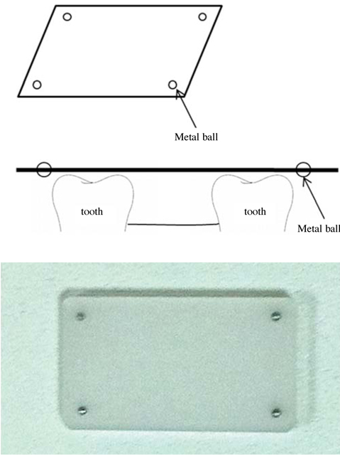
Fig. 1 Plastic plate for determination of a reference plane. For radiographic recognition, 4 metal balls (2 mm in diameter) are inserted in each of the 4 corners of the plate 2 mm away from the edge of the plate.

Fig. 2 Implant planning procedure using V-Implant software.
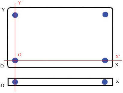
Fig. 3 The reference XOY-plane determined on the plastic plate to calculate the relative position and angulation of the virtual implant.
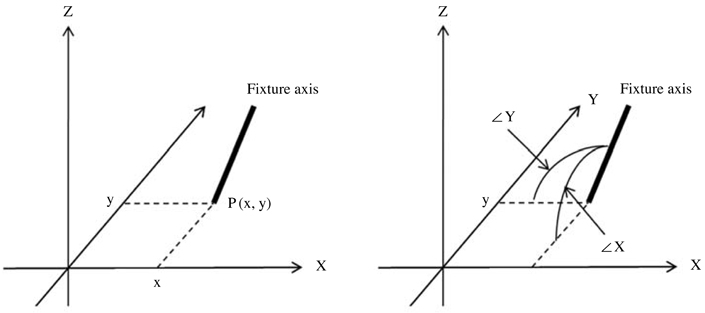
Fig. 4 These illustrations show the procedure for the determination of the position and angle of the virtual implant. The insertion point P (x, y) is determined by the crossing point between the axis of the virtual implant and the XOY-plane, and ∠X (Xθ) and ∠Y (Yθ) are defined as the degree from the X- and Y-axis, respectively.

Fig. 5 At the insertion points P (x, y), ∠X(Xθ) and ∠Y(Yθ) are calculated automatically by the software.

Fig. 6 A. The specially designed 5-axis drill machine. B. By moving and rotating the table of the machine, a guide hole is made reflecting the determined implant axis.
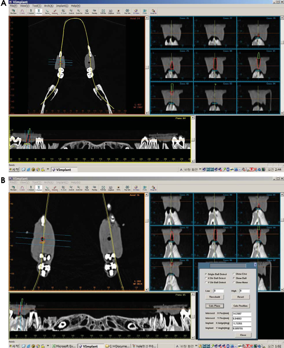
Fig. 7 A. The guide hole is shown as the black cylindrical area on the CT image. The virtual implant is placed into the guide hole on the software. B. After confirming the virtually positioned implants to be placed exactly into the guide hole, the position and the angulation of the central axis of the implant are calculated.
Reference
-
1. Branemark PI, Adell R, Breine U, Hansson BO, Lindstrom J, Ohlsson A. Intra-osseous anchorage of dental prostheses. I. Experimental studies. Scand J Plast Reconstr Surg. 1969. 3:81–100.
Article2. Israelson H, Plemons JM, Watkins P, Sory C. Barium-coated surgical stents and computer-assisted tomography in the preoperative assessment of dental implant patients. Int J Periodontics Restorative Dent. 1992. 12:52–61.3. Espinosa Marino J, Alvarez Arenal A, Pardo Ceballos A, Fernandez Vazquez JP, Ibaseta Diaz G. Fabrication of an implant radiologic-surgical stent for the partially edentulous patient. Quintessence Int. 1995. 26:111–114.4. Basten CH. The use of radiopaque templates for predictable implant placement. Quintessence Int. 1995. 26:609–612.5. Borrow JW, Smith JP. Stent marker materials for computerized tomograph-assisted implant planning. Int J Periodontics Restorative Dent. 1996. 16:60–67.6. Basten CH, Kois JC. The use of barium sulfate for implant templates. J Prosthet Dent. 1996. 76:451–454.
Article7. Weinberg LA, Kruger B. Three-dimensional guidance system for implant insertion: Part I. Implant Dent. 1998. 7:81–93.
Article8. Weinberg LA, Kruger B. Three-dimensional guidance system for implant insertion: Part II. Dual axes table-problem solving. Implant Dent. 1999. 8:255–264.9. Becker CM, Kaiser DA. Surgical guide for dental implant placement. J Prosthet Dent. 2000. 83:248–251.
Article10. Cucchiara R, Franchini F, Lamma A, Lamma E, Sansoni T, Sarti E. Enhancing implant surgery planning via computerized image processing. Int J Comput Dent. 2001. 4:9–24.11. Cucchiara R, Lamma E, Sansoni T. An image analysis approach for automatically re-orienteering CT images for dental implants. Comput Med Imaging Graph. 2004. 28:185–201.
Article12. Owings JR Jr. Virtual imaging guiding implant surgery. Compend Contin Educ Dent. 2003. 24:333–344.13. Marchack CB. An immediately loaded CAD/CAM-guided definitive prosthesis: a clinical report. J Prosthet Dent. 2005. 93:8–12.
Article14. Mupparapu M, Singer SR. Implant imaging for the dentist. J Can Dent Assoc. 2004. 70:32.15. Tardieu PB, Vrielinck L, Escolano E. Computer-assisted implant placement. A case report: treatment of the mandible. Int J Oral Maxillofac Implants. 2003. 18:599–604.16. Widmann G, Zangerl A, Keiler M, Stoffner R, Bale R, Puelacher W. Flapless implant surgery in the edentulous jaw based on three fixed intoral reference points and image-guided surgical templates: accuracy in human cadavers. Clin Oral Implants Res. 2010. 21:835–841.17. Widmann G, Keiler M, Zangerl A, Stoffner R, Longato S, Bale R, et al. Computer-assisted surgery in the edentulous jaw based on 3 fixed intraoral reference points. J Oral Maxillofac Surg. 2010. 68:1140–1147.
Article18. Pongrácz F, Bárdosi Z, Szabó L. Dentition planning for image-guided implantology. Int Congr Ser. 2004. 1268:1168–1173.
Article19. Koulechov K, Lueth T. A new metric for drill location for navigated control in navigated dental implantology. Int Congr Ser. 2004. 1268:1220–1225.
Article20. Parel SM, Triplett RG. Interactive imaging for implant planning, placement, and prosthesis construction. J Oral Maxillofac Surg. 2004. 62:9 suppl 2. 41–47.
Article21. Besimo CE, Lambrecht JT, Guindy JS. Accuracy of implant treatment planning utilizing template-guided reformatted computed tomography. Dentomaxillofac Radiol. 2000. 29:46–51.
Article22. Fortin T, Champleboux G, Lormee J, Coudert JL. Precise dental implant placement in bone using surgical guides in conjunction with medical imaging techniques. J Oral Implantol. 2000. 26:300–303.
Article23. Widmann G, Widmann R, Widmann E, Jaschke W, Bale RJ. In vitro accuracy of a novel registration and targeting technique for image-guided template production. Clin Oral Implants Res. 2005. 16:502–508.24. Brief J, Edinger D, Hassfeld S, Eggers G. Accuracy of image-guided implantology. Clin Oral Implants Res. 2005. 16:495–501.
Article25. Wanschitz F, Birkfellner W, Watzinger F, Schopper C, Patruta S, Kainberger F, et al. Evaluation of accuracy of computer-aided intraoperative positioning of endosseous oral implants in the edentulous mandible. Clin Oral Implants Res. 2002. 13:59–64.
Article26. Van Steenberghe D, Malevez C, Van Cleynenbreugel J, Bou Serhal C, Dhoore E, Schutyser F, et al. Accuracy of drilling guides for transfer from three-dimensional CT-based planning to placement of zygoma implants in human cadavers. Clin Oral Implants Res. 2003. 14:131–136.
Article
- Full Text Links
-
- Actions
-
Cited
- CITED
-
- Close
- Share
-
- Similar articles
-
- Surgical stent for dental implant using cone beam CT images
- An assessment of accuracy of half-guided implant surgery using implant surgical guide: A case report
- Current trends in dental implants
- The accuracy evaluation of digital surgical stents according to supported type
- Comparison of accuracy between free-hand and surgical guide implant placement among experienced and non-experienced dental implant practitioners: an in vitro study
