Primary Epithelial Ovarian Carcinoma with Gastric Metastasis Mimic Gastrointestinal Stromal Tumor
- Affiliations
-
- 1Department of Obstetrics and Gynecology, Chonnam National University Medical School, Gwangju, Korea. seokmo2001@hanmail.net
- 2Department of Pathology, Chonnam National University Medical School, Gwangju, Korea.
- 3Department of General Surgery, Chonnam National University Medical School, Gwangju, Korea.
Abstract
- Epithelial ovarian carcinoma rarely metastasizes to the parenchyma of the stomach. A 55-years-old woman presented with epigastric pain and a feeling of fullness for one month. A subsequent contrast-enhanced CT scan demonstrated a 4.5 x 4 cm submucosal mass with focal ulceration in the gastric antrum, and this finding was suggestive of GIST. After gastric antrectomy, the final pathology showed metastatic gastric tumor from a primary ovarian serous carcinoma. Because epithelial ovarian carcinoma is usually spread along the peritoneal surface, stomach involvement is rare. Furthermore, transmural gastric metastasis is very rare in a patient with primary ovarian carcinoma. Until now, there has been no reported case of stomach involvement at presentation in a patient with primary ovarian carcinoma. We present here a case of ovarian carcinoma with gastric metastasis that mimicked GIST.
MeSH Terms
Figure
-
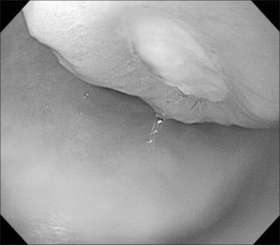
Fig. 1 Endoscopic view of the protruding tumor in the stomach with central ulceration.
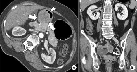
Fig. 2 The left decubitus CT image (A) shows a 4.5×4 cm extrinsic mass (arrows) smoothly compressing the posterior wall of the gastric antrum and abutting to the head of pancreas, which is suggestive of a gastric submucosal tumor such as gastrointestinal stromal tumor (GIST). A focal central ulceration (arrow head) is also noted. The coronal-reformatted CT image (B) demonstrates a 3 cm solid mass (curved arrow) in the pelvic cavity, corresponding to the right ovarian cancer, although it was considered as a metastatic lesion or synchronous small bowel GIST arising in the distal ileum at the time of imaging.
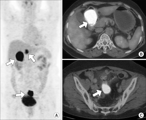
Fig. 3 The maximum intensity projection (MIP) reconstruction of the CT-based attenuation-corrected coronal PET image (A) shows multiple hypermetabolic lesions (max SUV, 22.3) in the abdominopelvic region (arrows). The axial fused PET/CT image (B and C) shows intense FDG uptake lesions (max SUV, 22.3) (arrows) in both the gastric antrum and the pelvic cavity abutting to the distal ileum, a finding that can mimic malignant gastric GIST with metastases or with a synchronous small bowel GIST arising from the distal ileum.
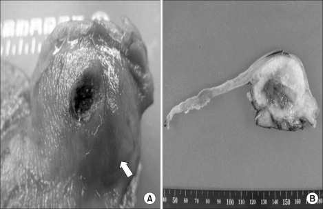
Fig. 4 Gross photograph of the stomach mass. In (A) the arrow points to the 4.5×4 cm sized submucosal mass of the stomach. (B) is the cut section of the stomach mass. The submucosal mass is shown below the normal mucosa.
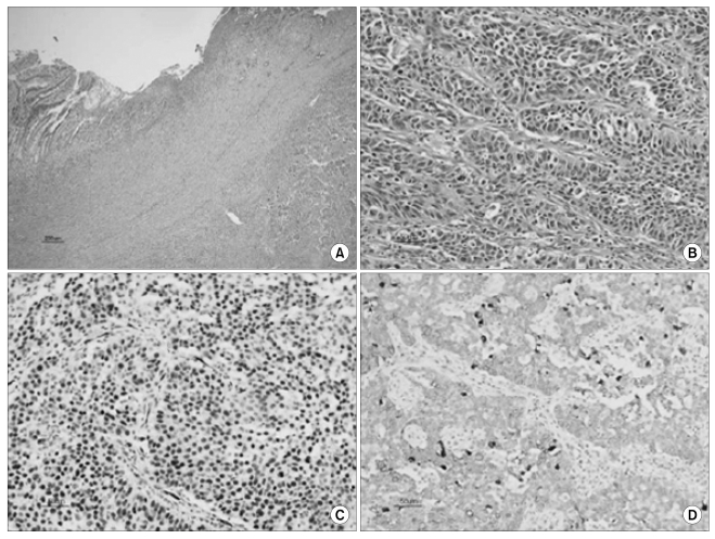
Fig. 5 The histologic and immunohistochemical findings. Low magnification shows an intramural tumor and the overlying ulcer (A, hematoxylin-eosin, ×40). This tumor is composed of irregular sheets of cells with high-grade nuclear atypia (B, hematoxylin-eosin, ×200). Immunohistochemically, the tumor cells are immunoreactive for Wilms' tumor-1 (C, ×200) and cytokeratin 7 (D, ×200).
Reference
-
1. Taylor RR, Phillips WS, O'Connor DM. Unusual intramural gastric metastasis of recurrent epithelial ovarian carcinoma. Gynecol Oncol. 1994; 55:152–155. PMID: 7959258.
Article2. Green LK. Hematogenous metastasis to the stomach. A review of 67 cases. Cancer. 1990; 65:1596–1600. PMID: 2311070.3. Nakatsuka S, Oji Y, Horiuchi T. Immunohistochemical detection of WT1 protein in a variety of cancer cells. Mod Pathol. 2006; 19:804–814. PMID: 16547468.
Article4. Tokunaga A, Nishi K, Matsukura N. Estrogen and progesterone receptors in gastric cancer. Cancer. 1986; 57:1376–1379. PMID: 3948119.
Article5. Berezowski K, Stastny JF, Kornstein MJ. Cytokeratins 7 and 20 and carcinoembryonic antigen in ovarian and colonic carcinoma. Mod Pathol. 1996; 9:426–429. PMID: 8729984.6. Loy TS, Calaluce RD, Keeney GL. Cytokeratin immunostaining in differentiating primary ovarian carcinoma from metastatic colonic adenocarcinoma. Mod Pathol. 1996; 9:1040–1044. PMID: 8933513.7. Wang NP, Zee S, Zarbo RJ. Coordinate expression of cytokeratins 7 and 20 defines unique subsets of carcinomas. Appl Immunohistochem. 1995; 3:99–107.
- Full Text Links
-
- Actions
-
Cited
- CITED
-
- Close
- Share
-
- Similar articles
-
- Gastric Metastasis from Ovarian Adenocarcinoma Presenting as a Submucosal Tumor without Ulceration
- Gastrointestinal Stromal Tumor with Extensive Lymphatic Metastasis: A Case Report
- A Case of Cerebral Metastsis Secondary to Primary Epithelial OvarianCarcinoma : in Complete Responder to Chemotherapy and Surgery
- A Case of Primary Peritoneal Carcinoma
- Immunohistochemical Studies for Differential Diagnosis between Primary and Metastatic OvarianEpithelial Tumors
