Ultrasonographic Diagnosis of Non-displaced Avulsion Fracture of the Acromion: A Case Report
- Affiliations
-
- 1Department of Rehabilitation Medicine, Maryknoll Medical Center, Busan, Korea.
- 2Department of Rehabilitation Medicine, Seoul National University College of Medicine, Seoul, Korea.
- 3Department of Rehabilitation Medicine, Seoul National University Boramae Medical Center, Seoul, Korea. ShiUk.Lee@gmail.com
- KMID: 2165651
- DOI: http://doi.org/10.5535/arm.2015.39.3.473
Abstract
- Avulsion fracture of the acromion is rare. It is difficult to diagnosis because there is little displacement and it occurs even without direct trauma. We experienced a case without direct trauma that was diagnosed with ultrasonography. A 55-year-old male patient visited our outpatient clinic with shoulder pain resulting from a significant stress at the trapezius muscle during lifting of a steel reinforcement. Simple radiography revealed a calcific deposit over the acromion rather than a fracture. Avulsion fracture was identified with ultrasonography. This is the first report demonstrating that ultrasonography has an advantage over radiographs in the diagnosis of an avulsion fracture of the acromion of the scapula.
Keyword
MeSH Terms
Figure
-
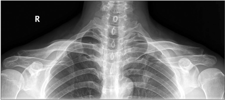
Fig. 1 The space between acromioclavicular joint is identical on both sides in the anterior-posterior view obtained with the both clavicles of the patient bearing weight. This finding suggests intact acromioclavicular ligament.
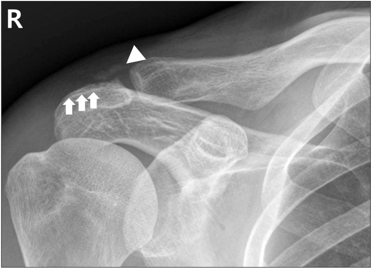
Fig. 2 Radiograph of shoulder in anterior-posterior view showed a small calcific deposit (arrowhead) and a possible fracture line (arrow) in the posterior area of the acromial process at right acromioclavicular joint. The possible fracture line was found after performing ultrasonography examination.
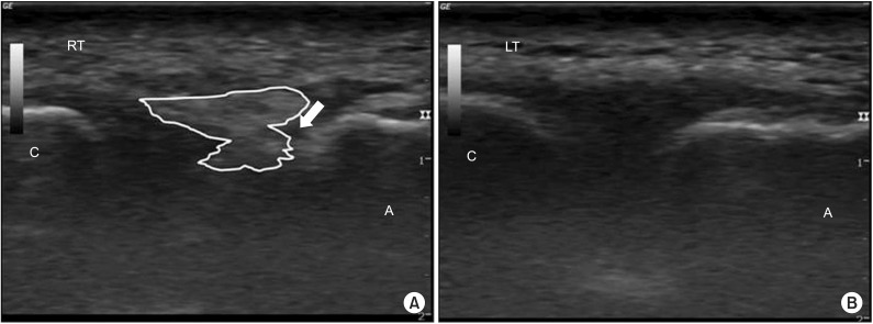
Fig. 3 Ultrasonographic image of acromioclavicular joint with oblique coronal view. (A) A minimally displaced extracapsular calcifications and defect (arrow) of the cortical surface at the acromion are shown. (B) Normal acromioclavicular joint on left side is shown. A, acromion; C, clavicle.
Reference
-
1. Rockwood CA, Beaty JH, Kasser JR. Rockwood and Wilkins' fractures in children. 7th ed. Philadelphia: Lippincott Williams & Wilkins;2010.2. Ada JR, Miller ME. Scapular fractures: analysis of 113 cases. Clin Orthop Relat Res. 1991; (269):174–180. PMID: 1864036.3. Goss TP. The scapula: coracoid, acromial, and avulsion fractures. Am J Orthop (Belle Mead NJ). 1996; 25:106–115. PMID: 8640380.4. Kuhn JE, Blasier RB, Carpenter JE. Fractures of the acromion process: a proposed classification system. J Orthop Trauma. 1994; 8:6–13. PMID: 8169698.
Article5. Patten RM, Mack LA, Wang KY, Lingel J. Nondisplaced fractures of the greater tuberosity of the humerus: sonographic detection. Radiology. 1992; 182:201–204. PMID: 1727282.
Article6. Senall JA, Failla JM, Bouffard JA, van Holsbeeck M. Ultrasound for the early diagnosis of clinically suspected scaphoid fracture. J Hand Surg Am. 2004; 29:400–405. PMID: 15140480.
Article7. Heyse-Moore GH, Stoker DJ. Avulsion fractures of the scapula. Skeletal Radiol. 1982; 9:27–32. PMID: 7157014.
Article8. Rask MR, Steinberg LH. Fracture of the acromion caused by muscle forces: a case report. J Bone Joint Surg Am. 1978; 60:1146–1147. PMID: 721873.
Article9. Grechenig W, Clement H, Schatz B, Klein A, Grechenig M. Die sonographische frakturdiagnostik: eine experimentelle studie. Biomed Tech (Berl). 1997; 42:138–145. PMID: 9272995.
- Full Text Links
-
- Actions
-
Cited
- CITED
-
- Close
- Share
-
- Similar articles
-
- Isolated Avulsion Fracture of the Superior Border of the Scapula: A Case Report
- Sequela of Untreated Avulsion Fracutre of Ischial Tuberosity: Report of Two Cases
- Displaced fracture of the base of the second metacarpal into velar side
- Acute Displaced Fracture of Lateral Acromion after Reverse Shoulder Arthroplasty: A Case Report and Surgical Technique
- Avulsion Fracture of Calcaneal Tubercle Treated with Cannulated Cancellous Screws and Wire: Surgical Technique
