Primary Fallopian Tube Carcinoma Diagnosed with Endoscopic Ultrasound Elastography with Fine Needle Biopsy
- Affiliations
-
- 1Institution for Digestive Research, Digestive Disease Center, Department of Internal Medicine, Soonchunhyang University Hospital, Soonchunhyang University College of Medicine, Seoul, Korea. iman0825@schmc.ac.kr
- 2Department of Obstetrics and Gynecology, Soonchunhyang University College of Medicine, Seoul, Korea.
- 3Department of Pathology, Soonchunhyang University College of Medicine, Seoul, Korea.
- KMID: 2165382
- DOI: http://doi.org/10.5946/ce.2014.47.5.464
Abstract
- Primary fallopian tube carcinoma (PFTC) is a rare gynecological cancer that is very difficult to diagnose preoperatively. Here, we report the case of a 66-year-old female patient with PFTC that was diagnosed preoperatively on the basis of the characteristic features on endoscopic ultrasound (EUS) elastography and fine needle biopsy (FNB). EUS showed a sausage-shaped hypoechoic mass, 8 cm in size, with irregular margins and heterogeneous internal echoes extending to both adnexa. EUS elastography revealed that the mass had a blue color pattern, representing hard stiffness, and a heterogeneous green/red color pattern distributed outside the tumor, representing intermediate stiffness. Histopathologic analysis of the FNB and operative specimens confirmed the diagnosis of fallopian tube carcinoma. This is the first reported case of a combined EUS elastography and FNB of an adnexal mass leading to a preoperative diagnosis of fallopian tube carcinoma.
Keyword
MeSH Terms
Figure
-
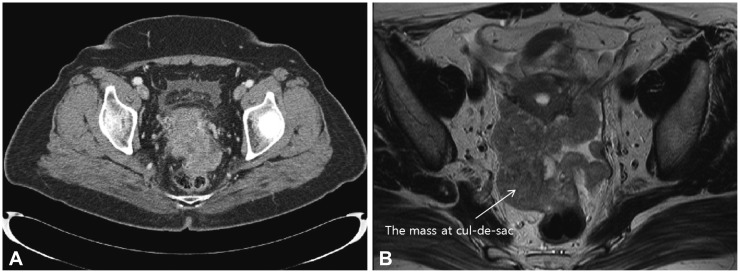
Fig. 1 Computed tomography (CT) and magnetic resonance imaging (MRI) of the adnexal mass. (A) An about 8-cm, heterogeneous enhancing mass with irregular shape, involving from the uterine cervix to the uterine fundus, was detected by using CT. (B) The mass at cul-de-sac (white arrow) has directly invaded to the rectum on MRI.
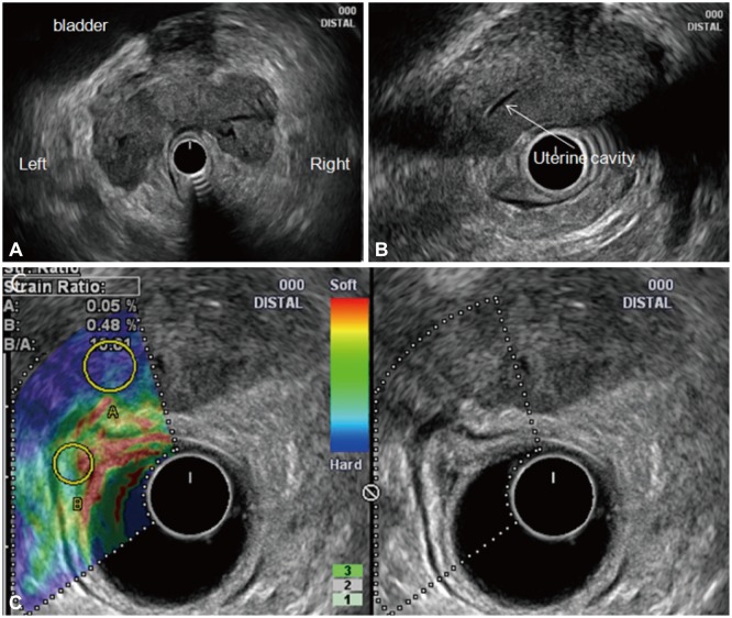
Fig. 2 Endoscopic ultrasound elastography of the adnexal mass. (A) A sausage-shaped hypoechoic mass, 8 cm in size, with irregular margins and heterogeneous internal echoes extending to both adnexa and that had obliterated the lumen of both fallopian tubes was detected. (B) The mass had invaded the right side of the uterine body, making it difficult to delineate the borderline between the lesion and the uterus. (C) Elastographic image of the left side of the dual image showing the blue color pattern (representing hard stiffness) of the mass and heterogeneous green/red coloration (representing intermediate stiffness) outside the tumor.
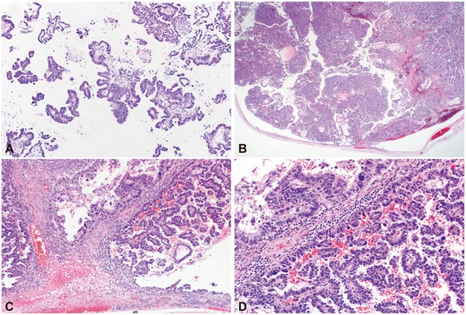
Fig. 3 Histopathologic analyses of needle biopsy and operative tissue specimens. (A) The ProCore needle biopsy specimen revealed papillary growing tumor cells with hyperchromatic nuclei and frequent mitoses, suggestive of primary or metastatic papillary adenocarcinoma of the pelvic cavity (H&E stain, ×100). (B) The operative specimen revealed a tumor occupying the lumen of the right residual fallopian tube that had extended to the pelvic cavity (H&E stain, ×12.5). (C) Operative specimen showing the tumor arising from the luminal epithelium with a papillary growth pattern (H&E stain, ×100). (D) Operative specimen showing that the tumor consisted of a fibrovascular core lined by a single layer of tumor cells with occasional pleomorphic cells and frequent atypical mitosis (H&E stain, ×200). These findings are similar to those of the endoscopic ultrasound-guided biopsy.
Reference
-
1. Semrad N, Watring W, Fu YS, Hallatt J, Ryoo M, Lagasse L. Fallopian tube adenocarcinoma: common extraperitoneal recurrence. Gynecol Oncol. 1986; 24:230–235. PMID: 3710267.
Article2. Pectasides D, Pectasides E, Economopoulos T. Fallopian tube carcinoma: a review. Oncologist. 2006; 11:902–912. PMID: 16951394.
Article3. Itoh A, Ueno E, Tohno E, et al. Breast disease: clinical application of US elastography for diagnosis. Radiology. 2006; 239:341–350. PMID: 16484352.
Article4. Cochlin DL, Ganatra RH, Griffiths DF. Elastography in the detection of prostatic cancer. Clin Radiol. 2002; 57:1014–1020. PMID: 12409113.
Article5. Krouskop TA, Wheeler TM, Kallel F, Garra BS, Hall T. Elastic moduli of breast and prostate tissues under compression. Ultrason Imaging. 1998; 20:260–274. PMID: 10197347.
Article6. Giovannini M, Thomas B, Erwan B, et al. Endoscopic ultrasound elastography for evaluation of lymph nodes and pancreatic masses: a multicenter study. World J Gastroenterol. 2009; 15:1587–1593. PMID: 19340900.
Article7. Gheorghe L, Gheorghe C, Cotruta B, Carabela A. CT aspects of gastrointestinal stromal tumors: adding EUS and EUS elastography to the diagnostic tools. J Gastrointestin Liver Dis. 2007; 16:346–347. PMID: 17925934.8. Iglesias-Garcia J, Larino-Noia J, Abdulkader I, Forteza J, Dominguez-Munoz JE. Quantitative endoscopic ultrasound elastography: an accurate method for the differentiation of solid pancreatic masses. Gastroenterology. 2010; 139:1172–1180. PMID: 20600020.
Article9. Rose PG, Piver MS, Tsukada Y. Fallopian tube cancer. The Roswell Park experience. Cancer. 1990; 66:2661–2667. PMID: 2249208.
Article10. Rosen A, Klein M, Lahousen M, Graf AH, Rainer A, Vavra N. Austrian Cooperative Study Group for Fallopian Tube Carcinoma. Primary carcinoma of the fallopian tube: a retrospective analysis of 115 patients. Br J Cancer. 1993; 68:605–609. PMID: 8353051.11. Peters WA 3rd, Andersen WA, Hopkins MP, Kumar NB, Morley GW. Prognostic features of carcinoma of the fallopian tube. Obstet Gynecol. 1988; 71:757–762. PMID: 3357664.
Article12. Alvarado-Cabrero I, Young RH, Vamvakas EC, Scully RE. Carcinoma of the fallopian tube: a clinicopathological study of 105 cases with observations on staging and prognostic factors. Gynecol Oncol. 1999; 72:367–379. PMID: 10053109.
Article13. Gadducci A, Landoni F, Sartori E, et al. Analysis of treatment failures and survival of patients with fallopian tube carcinoma: a cooperation task force (CTF) study. Gynecol Oncol. 2001; 81:150–159. PMID: 11330942.
Article14. Ajjimakorn S, Bhamarapravati Y, Israngura N. Ultrasound appearance of fallopian tube carcinoma. J Clin Ultrasound. 1988; 16:516–518. PMID: 3152451.
Article15. Kol S, Gal D, Friedman M, Paldi E. Preoperative diagnosis of fallopian tube carcinoma by transvaginal sonography and CA-125. Gynecol Oncol. 1990; 37:129–131. PMID: 2182403.
Article16. Subramanyam BR, Raghavendra BN, Whalen CA, Yee J. Ultrasonic features of fallopian tube carcinoma. J Ultrasound Med. 1984; 3:391–393. PMID: 6384545.
Article17. Ophir J, Céspedes I, Ponnekanti H, Yazdi Y, Li X. Elastography: a quantitative method for imaging the elasticity of biological tissues. Ultrason Imaging. 1991; 13:111–134. PMID: 1858217.
Article18. Lee TH, Cha SW, Cho YD. EUS elastography: advances in diagnostic EUS of the pancreas. Korean J Radiol. 2012; 13(Suppl 1):S12–S16. PMID: 22563282.
Article
- Full Text Links
-
- Actions
-
Cited
- CITED
-
- Close
- Share
-
- Similar articles
-
- Two Cases of Primary Carcinoma in the Fallopian Tube
- Two Cases of Primary Carcinoma of the Fallopian Tube
- Primary Squamous Cell Carcinoma of the Gallbladder Diagnosed by Endoscopic Ultrasound-Guided Fine Needle Biopsy
- Endoscopic Ultrasound-Fine Needle Aspiration versus Core Biopsy for the Diagnosis of Subepithelial Tumors
- Fine-Needle Biopsy: Should This Be the First Choice in Endoscopic Ultrasound-Guided Tissue Acquisition?
