Mechanisms of Biliary Plastic Stent Occlusion and Efforts at Prevention
- Affiliations
-
- 1Digestive Disease Center, CHA Bundang Medical Center, CHA University, Seongnam, Korea.
- 2Division of Gastroenterology and Hepatology, Indiana University School of Medicine, Indianapolis, IN, USA. glehman@iu.edu
- KMID: 2165037
- DOI: http://doi.org/10.5946/ce.2016.024
Abstract
- Biliary stenting via endoscopic retrograde cholangiopancreatography has greatly improved the quality of patient care over the last 30 years. Plastic stent occlusion limits the life span of such stents. Attempts to improve plastic stent patency duration have mostly failed. Metal stents (self-expandable metal stents [SEMSs]) have therefore replaced plastic stents, especially for malignant biliary strictures. SEMS are at least 10 times more expensive than plastic stents. In this focused review, we will discuss basic mechanisms of plastic stent occlusion, along with a systematic summary of previous efforts and related studies to improve stent patency and potential new techniques to overcome existing limitations.
Keyword
MeSH Terms
Figure
-

Fig. 1. (A-E) Retrieved biliary plastic stents with the inner layer exposed by longitudinal cutting. The dark greenish biofilms do not contribute significantly to increased thickness of the occluded inner layer. Stent occlusion is mostly caused by debris, sludge, and food components.
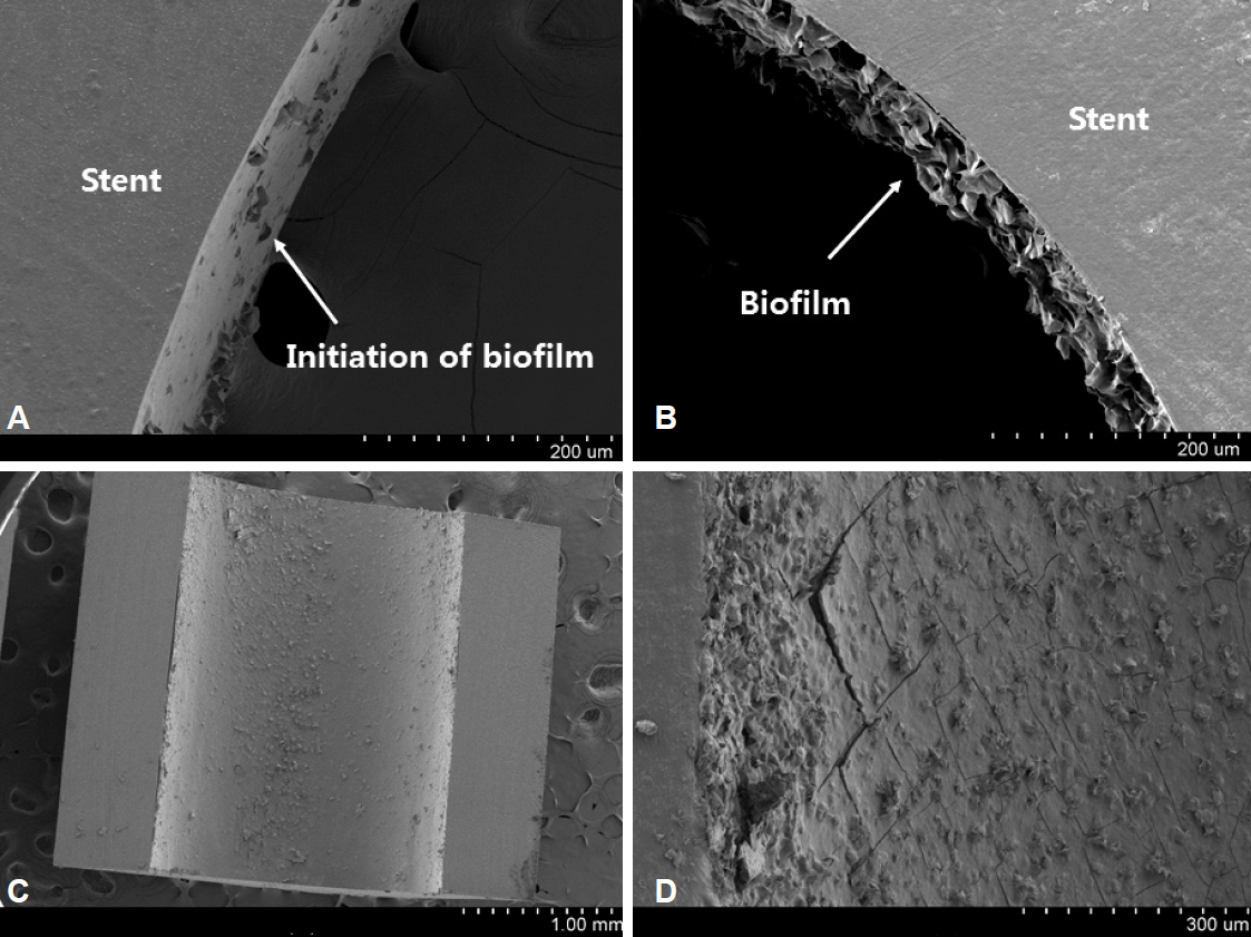
Fig. 2. Scanning electron microscopy (SEM) examination of stent occlusion. SEM images of the inner surface of a stent retrieved at about 4 weeks. The biofilm starts to appear on the inner surface of the stent (A, ×250). The biofilm itself becomes gradually thicker relatively evenly (B, ×250; C, ×30). The surface becomes more solid and cracked in some areas (D, ×150).

Fig. 3. Scanning electron microscopy (SEM) examination of stent occlusion. SEM images of the inner surface of a stent retrieved at about 8 weeks. Sludge covers the biofilm and narrows the inner diameter of the stent (A, ×30; B, ×150). The biofilm is exposed in some areas that are not covered by sludge (C, ×150). Debris, presumably derived from other causes even before the formation of the biofilm, is attached to the inner layer of the stent (D, ×100).
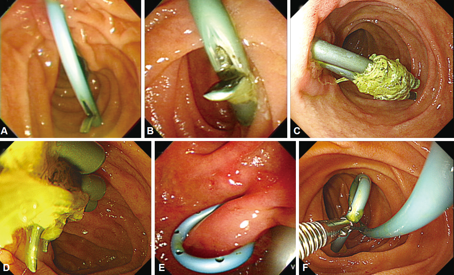
Fig. 4. Factors causing plastic biliary stent malfunction. (A, B) Distal ends of the straight stents are touching the duodenal wall as a result of partial distal migration. This may lead to flow disturbance and sludge accumulation, causing occlusion. (C, D) The straight stents have not migrated, but dietary fibers are clinging to the side flaps, causing stent occlusion. (E, F) Distal ends of the pigtail stents are touching the duodenal wall or partially migrated, but the side holes still allow bile flow.
Cited by 1 articles
-
Relief of Obstruction in the Management of Pancreatic Cancer
Chang-Il Kwon
Korean J Gastroenterol. 2019;74(2):69-80. doi: 10.4166/kjg.2019.74.2.69.
Reference
-
1. Ballinger AB, McHugh M, Catnach SM, Alstead EM, Clark ML. Symptom relief and quality of life after stenting for malignant bile duct obstruction. Gut. 1994; 35:467–470.
Article2. Faigel DO. Preventing biliary stent occlusion. Gastrointest Endosc. 2000; 51:104–107.
Article3. Raijman I. Biliary and pancreatic stents. Gastrointest Endosc Clin N Am. 2003; 13:561–592.
Article4. Soehendra N, Reynders-Frederix V. Palliative bile duct drainage: a new endoscopic method of introducing a transpapillary drain. Endoscopy. 1980; 12:8–11.5. Speer AG, Cotton PB, Russell RC, et al. Randomised trial of endoscopic versus percutaneous stent insertion in malignant obstructive jaundice. Lancet. 1987; 2:57–62.
Article6. Libby ED, Leung JW. Prevention of biliary stent clogging: a clinical review. Am J Gastroenterol. 1996; 91:1301–1308.7. Leung JW, Liu Y, Chan RC, et al. Early attachment of anaerobic bacteria may play an important role in biliary stent blockage. Gastrointest Endosc. 2000; 52:725–729.
Article8. van Berkel AM, van Marle J, Groen AK, Bruno MJ. Mechanisms of biliary stent clogging: confocal laser scanning and scanning electron microscopy. Endoscopy. 2005; 37:729–734.
Article9. Guaglianone E, Cardines R, Vuotto C, et al. Microbial biofilms associated with biliary stent clogging. FEMS Immunol Med Microbiol. 2010; 59:410–420.
Article10. Groen AK, Out T, Huibregtse K, Delzenne B, Hoek FJ, Tytgat GN. Characterization of the content of occluded biliary endoprostheses. Endoscopy. 1987; 19:57–59.
Article11. Speer AG, Cotton PB, Rode J, et al. Biliary stent blockage with bacterial biofilm. A light and electron microscopy study. Ann Intern Med. 1988; 108:546–553.12. Leung JW, Ling TK, Kung JL, Vallance-Owen J. The role of bacteria in the blockage of biliary stents. Gastrointest Endosc. 1988; 34:19–22.
Article13. Moesch C, Sautereau D, Cessot F, et al. Physicochemical and bacteriological analysis of the contents of occluded biliary endoprostheses. Hepatology. 1991; 14:1142–1146.
Article14. An YH, Friedman RJ. Concise review of mechanisms of bacterial adhesion to biomaterial surfaces. J Biomed Mater Res. 1998; 43:338–348.
Article15. Chan FK, Suen M, Li JY, Sung JJ. Bile immunoglobulins and blockage of biliary endoprosthesis: an immunohistochemical study. Biomed Pharmacother. 1998; 52:403–407.
Article16. Yu JL, Andersson R, Ljungh A. Protein adsorption and bacterial adhesion to biliary stent materials. J Surg Res. 1996; 62:69–73.
Article17. Yu JL, Andersson R, Wang LQ, Bengmark S, Ljungh A. Fibronectin on the surface of biliary drain materials: a role in bacterial adherence. J Surg Res. 1995; 59:596–600.18. Donelli G, Guaglianone E, Di Rosa R, Fiocca F, Basoli A. Plastic biliary stent occlusion: factors involved and possible preventive approaches. Clin Med Res. 2007; 5:53–60.
Article19. Speer AG, Cotton PB, MacRae KD. Endoscopic management of malignant biliary obstruction: stents of 10 French gauge are preferable to stents of 8 French gauge. Gastrointest Endosc. 1988; 34:412–417.
Article20. ASGE Technology Assessment Committee, Pfau PR, Pleskow DK, et al. Pancreatic and biliary stents. Gastrointest Endosc. 2013; 77:319–327.
Article21. Gilbert DA, DiMarino AJ Jr, Jensen DM, et al. Status evaluation: biliary stents. American Society for Gastrointestinal Endoscopy. Technology Assessment Committee. Gastrointest Endosc. 1992; 38:750–752.22. Kadakia SC, Starnes E. Comparison of 10 French gauge stent with 11.5 French gauge stent in patients with biliary tract diseases. Gastrointest Endosc. 1992; 38:454–459.
Article23. Wagh MS, de Bellis M, Fogel EL, et al. Multicenter randomized trial of 10-French versus 11.5-French plastic stents for malignant biliary obstruction. Diagn Ther Endosc. 2013; 2013:891915.
Article24. van Berkel AM, Boland C, Redekop WK, et al. A prospective randomized trial of Teflon versus polyethylene stents for distal malignant biliary obstruction. Endoscopy. 1998; 30:681–686.
Article25. England RE, Martin DF, Morris J, et al. A prospective randomised multicentre trial comparing 10 Fr Teflon Tannenbaum stents with 10 Fr polyethylene Cotton-Leung stents in patients with malignant common duct strictures. Gut. 2000; 46:395–400.
Article26. Terruzzi V, Comin U, De Grazia F, et al. Prospective randomized trial comparing Tannenbaum Teflon and standard polyethylene stents in distal malignant biliary stenosis. Gastrointest Endosc. 2000; 51:23–27.
Article27. Cheon YK, Oh HC, Cho YD, Lee TY, Shim CS. New 10F soft and pliable polyurethane stents decrease the migration rate compared with conventional 10F polyethylene stents in hilar biliary obstruction: results of a pilot study. Gastrointest Endosc. 2012; 75:790–797.
Article28. Motte S, Deviere J, Dumonceau JM, Serruys E, Thys JP, Cremer M. Risk factors for septicemia following endoscopic biliary stenting. Gastroenterology. 1991; 101:1374–1381.
Article29. Shepherd HA, Royle G, Ross AP, Diba A, Arthur M, Colin-Jones D. Endoscopic biliary endoprosthesis in the palliation of malignant obstruction of the distal common bile duct: a randomized trial. Br J Surg. 1988; 75:1166–1168.
Article30. Schmassmann A, von Gunten E, Knuchel J, Scheurer U, Fehr HF, Halter F. Wallstents versus plastic stents in malignant biliary obstruction: effects of stent patency of the first and second stent on patient compliance and survival. Am J Gastroenterol. 1996; 91:654–659.31. Kwon CI, Ko KH, Hahm KB, Kang DH. Functional self-expandable metal stents in biliary obstruction. Clin Endosc. 2013; 46:515–521.
Article32. Galandi D, Schwarzer G, Bassler D, Allgaier HP. Ursodeoxycholic acid and/or antibiotics for prevention of biliary stent occlusion. Cochrane Database Syst Rev. 2002. (3):CD003043.
Article33. Browne S, Schmalz M, Geenen J, Venu R, Johnson GK. A comparison of biliary and pancreatic stent occlusion in antibiotic-coated vs. conventional stents. Gastrointest Endosc. 1990; 36:206.34. Gwon DI, Lee SS, Kim EY. Cefotaxime-eluting covered self-expandable stents in a canine biliary model: scanning electron microscopic study of biofilm formation. Acta Radiol. 2012; 53:1127–1132.
Article35. Weickert U, Wiesend F, Subkowski T, Eickhoff A, Reiss G. Optimizing biliary stent patency by coating with hydrophobin alone or hydrophobin and antibiotics or heparin: an in vitro proof of principle study. Adv Med Sci. 2011; 56:138–144.
Article36. Dowidar N, Kolmos HJ, Matzen P. Experimental clogging of biliary endoprostheses. Role of bacteria, endoprosthesis material, and design. Scand J Gastroenterol. 1992; 27:77–80.
Article37. Jansen B, Goodman LP, Ruiten D. Bacterial adherence to hydrophilic polymer-coated polyurethane stents. Gastrointest Endosc. 1993; 39:670–673.
Article38. Sung JY, Shaffer EA, Lam K, Rususka I, Costerton JW. Hydrophobic bile salt inhibits bacterial adhesion on biliary stent material. Dig Dis Sci. 1994; 39:999–1006.
Article39. John SF, Hillier VF, Handley PS, Derrick MR. Adhesion of staphylococci to polyurethane and hydrogel-coated polyurethane catheters assayed by an improved radiolabelling technique. J Med Microbiol. 1995; 43:133–140.
Article40. McAllister EW, Carey LC, Brady PG, Heller R, Kovacs SG. The role of polymeric surface smoothness of biliary stents in bacterial adherence, biofilm deposition, and stent occlusion. Gastrointest Endosc. 1993; 39:422–425.
Article41. Hoffman BJ, Cunningham JT, Marsh WH, O’Brien JJ, Watson J. An in vitro comparison of biofilm formation on various biliary stent materials. Gastrointest Endosc. 1994; 40:581–583.
Article42. van Berkel AM, Bruno MJ, Bergman JJ, van Deventer SJ, Tytgat GN, Huibregtse K. A prospective randomized study of hydrophilic polymer-coated polyurethane versus polyethylene stents in distal malignant biliary obstruction. Endoscopy. 2003; 35:478–482.
Article43. Costamagna G, Mutignani M, Rotondano G, et al. Hydrophilic hydromer-coated polyurethane stents versus uncoated stents in malignant biliary obstruction: a randomized trial. Gastrointest Endosc. 2000; 51:8–11.
Article44. Leung JW, Lau GT, Sung JJ, Costerton JW. Decreased bacterial adherence to silver-coated stent material: an in vitro study. Gastrointest Endosc. 1992; 38:338–340.
Article45. Yang J, Linghu E, Wang Y. Development of antibacterial plastic biliary stent coated with nano-silver. Zhongguo Yi Liao Qi Xie Za Zhi. 2011; 35:352–355. 360. Chinese.46. Coene PP, Groen AK, Cheng J, Out MM, Tytgat GN, Huibregtse K. Clogging of biliary endoprostheses: a new perspective. Gut. 1990; 31:913–917.
Article47. Rey JF, Maupetit P, Greff M. Experimental study of biliary endoprosthesis efficiency. Endoscopy. 1985; 17:145–148.
Article48. Seitz U, Vadeyar H, Soehendra N. Prolonged patency with a new-design Teflon biliary prosthesis. Endoscopy. 1994; 26:478–482.
Article49. Binmoeller KF, Seitz U, Seifert H, Thonke F, Sikka S, Soehendra N. The Tannenbaum stent: a new plastic biliary stent without side holes. Am J Gastroenterol. 1995; 90:1764–1768.50. Dua KS, Reddy ND, Rao VG, Banerjee R, Medda B, Lang I. Impact of reducing duodenobiliary reflux on biliary stent patency: an in vitro evaluation and a prospective randomized clinical trial that used a biliary stent with an antireflux valve. Gastrointest Endosc. 2007; 65:819–828.
Article51. Leong QW, Shen ML, Au KW, et al. A prospective, randomized study of the patency period of the plastic antireflux biliary stent: an interim analysis. Gastrointest Endosc. 2016; 83:387–393.
Article52. Lee YN, Moon JH, Choi HJ, et al. Effectiveness of a newly designed antireflux valve metal stent to reduce duodenobiliary reflux in patients with unresectable distal malignant biliary obstruction: a randomized, controlled pilot study (with videos). Gastrointest Endosc. 2016; 83:404–412.
Article53. Hu B, Wang TT, Wu J, Shi ZM, Gao DJ, Pan YM. Antireflux stents to reduce the risk of cholangitis in patients with malignant biliary strictures: a randomized trial. Endoscopy. 2014; 46:120–126.
Article54. Kim DU, Kwon CI, Kang DH, Ko KH, Hong SP. New antireflux self-expandable metal stent for malignant lower biliary obstruction: in vitro and in vivo preliminary study. Dig Endosc. 2013; 25:60–66.55. Georghegan JG, Branch MS, Costerton JW, Pappas TN, Cotton PB. Placement of biliary stents above the sphincter of Oddi prolongs stent patency in dogs. Gut. 1991; 32:A1232.56. Pedersen FM, Lassen AT, Schaffalitzky de Muckadell OB. Randomized trial of stent placed above and across the sphincter of Oddi in malignant bile duct obstruction. Gastrointest Endosc. 1998; 48:574–579.
Article57. Cho JN, Han J, Kim HG, et al. Prospective randomized trial comparing covered metal stent placed above and across the sphincter of Oddi in malignant biliary obstruction. Gastrointest Endosc. 2013; 77(5 Suppl):AB139–AB140.
- Full Text Links
-
- Actions
-
Cited
- CITED
-
- Close
- Share
-
- Similar articles
-
- Stent-stone Complex Developed after Long-term Plastic Biliary Stent Placement
- A Comparative Study on the Efficacy of Covered Metal Stent and Plastic Stent in Malignant Biliary Obstruction
- Delayed Hemobilia Caused by Penetration of Biliary Plastic Stent into Portal Vein
- Endoscopic Reintervention for Recurrence of Malignant Biliary Obstruction: Developing the Best Strategy
- Articulated percutaneous plastic biliary stents: How to do it
