Identification of Outer Membrane Vesicles Derived from Orientia tsutsugamushi
- Affiliations
-
- 1Translation Research Center, Inha University School of Medicine, Incheon, Korea.
- 2Department of Internal Medicine, Inha University School of Medicine, Incheon, Korea. ljinsoo@inha.ac.kr
- 3Department of Microbiology, Inha University School of Medicine, Incheon, Korea.
- KMID: 2164469
- DOI: http://doi.org/10.3346/jkms.2015.30.7.866
Abstract
- Orientia tsutsugamushi, a causative pathogen of Scrub typhus, is a gram-negative intracellular bacterium. Outer membrane vesicles (OMVs) are produced from the membrane of bacteria and play many roles related to the survival of the pathogen. However, there have been no reports confirming whether O. tsutsugamushi indeed produce OMVs. O. tsutsugamushi boryong was cultured in ECV-304 cells for the purification of OMVs. Western blot analysis and immunoenrichment using anti-O. tsutsugamushi monoclonal antibody and electron microscopy were employed for identification and characterization of OMVs. We confirm the presence of OMVs derived from O. tsutsugamushi, and also found that those OMVs contain a major surface antigen of 56-kDa protein and variant immunogenic antigens.
MeSH Terms
-
Antibodies, Monoclonal/*immunology
Antigens, Bacterial/*immunology
Antigens, Surface/*immunology
Cell Line
Cell Membrane/immunology
Humans
Microscopy, Electron
Orientia tsutsugamushi/*immunology/metabolism
Scrub Typhus/diagnosis/microbiology
Secretory Vesicles/*immunology
Antibodies, Monoclonal
Antigens, Bacterial
Antigens, Surface
Figure
-
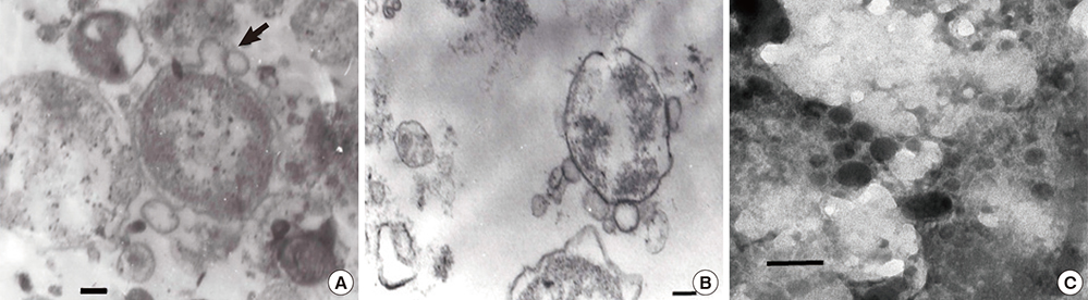
Fig. 1 Transmission electron micrographs of outer membrane vesicles of O. tsutsugamushi. (A) Blebs of vesicles (arrow) before they are liberated from O. tsutsugamushi (arrow). (B) Microvesicles are shown on the surface of purified O. tsutsugamushi. (C) Clusters of purified microvesicles are observed. They vary in size ranging from 50 to 150 nm and are made of monolayer membranes. Scale bars indicate 0.1 µm.

Fig. 2 Western blot analysis of purified and enriched OMVs derived from O. tsutsugamushi. (A) Blots were revealed with FS15 monoclonal antibody against OMVs and had similar patterns with blots of bacterial lysates. (B) Immunoblot of OMVs with polyclonal antibody showed a prominent 56-kDa protein band. (C) OMVs immunoenriched with FS15 monoclonal antibody revealed major surface antigens. ECV304, lysates of host cells; OT, lysates of purified O. tsutsugamushi; OMVs, outer membrane vesicles; FS15, mouse monoclonal antibody of O. tsutsugamushi; poly Ab, human polyclonal antibody purified from scrub typhus patient`s serum; IP, immunoprecipitation; IB, Immuno blot.
Reference
-
1. Tamura A, Ohashi N, Urakami H, Miyamura S. Classification of Rickettsia tsutsugamushi in a new genus, Orientia gen. nov., as Orientia tsutsugamushi comb. nov. Int J Syst Bacteriol. 1995; 45:589–591.2. Chung MH, Lee JS, Baek JH, Kim M, Kang JS. Persistence of Orientia tsutsugamushi in humans. J Korean Med Sci. 2012; 27:231–235.3. Seong SY, Choi MS, Kim IS. Orientia tsutsugamushi infection: overview and immune responses. Microbes Infect. 2001; 3:11–21.4. Korean Centers for Diseases Control and Prevention. Current status of selected infectious diseases. Public Health Wkly Rep. 2014; 27:585–591.5. Collins BS. Gram-negative outer membrane vesicles in vaccine development. Discov Med. 2011; 12:7–15.6. Kuehn MJ, Kesty NC. Bacterial outer membrane vesicles and the host-pathogen interaction. Genes Dev. 2005; 19:2645–2655.7. Kulp A, Kuehn MJ. Biological functions and biogenesis of secreted bacterial outer membrane vesicles. Annu Rev Microbiol. 2010; 64:163–184.8. Garcia-del Portillo F, Stein MA, Finlay BB. Release of lipopolysaccharide from intracellular compartments containing Salmonella typhimurium to vesicles of the host epithelial cell. Infect Immun. 1997; 65:24–34.9. Anthony LD, Burke RD, Nano FE. Growth of Francisella spp. in rodent macrophages. Infect Immun. 1991; 59:3291–3296.10. Stirling P, Richmond SJ. Production of outer membrane blebs during chlamydial replication. FEMS Microbiol Lett. 1980; 9:103–105.11. Tamura A, Urakami H, Tsuruhara T. Purification of Rickettsia tsutsugamushi by Percoll density gradient centrifugation. Microbiol Immunol. 1982; 26:321–328.12. Lee EY, Bang JY, Park GW, Choi DS, Kang JS, Kim HJ, Park KS, Lee JO, Kim YK, Kwon KH, et al. Global proteomic profiling of native outer membrane vesicles derived from Escherichia coli. Proteomics. 2007; 7:3143–3153.13. Schwechheimer C, Sullivan CJ, Kuehn MJ. Envelope control of outer membrane vesicle production in Gram-negative bacteria. Biochemistry. 2013; 52:3031–3040.14. Tamura A, Ohashi N, Urakami H, Takahashi K, Oyanagi M. Analysis of polypeptide composition and antigenic components of Rickettsia tsutsugamushi by polyacrylamide gel electrophoresis and immunoblotting. Infect Immun. 1985; 48:671–675.15. Seong SY, Huh MS, Jang WJ, Park SG, Kim JG, Woo SG, Choi MS, Kim IS, Chang WH. Induction of homologous immune response to Rickettsia tsutsugamushi Boryong with a partial 56-kilodalton recombinant antigen fused with the maltose-binding protein MBP-Bor56. Infect Immun. 1997; 65:1541–1545.16. Seong SY, Kim HR, Huh MS, Park SG, Kang JS, Han TH, Choi MS, Chang WH, Kim IS. Induction of neutralizing antibody in mice by immunization with recombinant 56 kDa protein of Orientia tsutsugamushi. Vaccine. 1997; 15:1741–1747.17. Frohlich K, Hua Z, Wang J, Shen L. Isolation of Chlamydia trachomatis and membrane vesicles derived from host and bacteria. J Microbiol Methods. 2012; 91:222–230.18. Lee DH, Kim SH, Kang W, Choi YS, Lee SH, Lee SR, You S, Lee HK, Chang KT, Shin EC. Adjuvant effect of bacterial outer membrane vesicles with penta-acylated lipopolysaccharide on antigen-specific T cell priming. Vaccine. 2011; 29:8293–8301.19. Bosma T, Kanninga R, Neef J, Audouy SA, van Roosmalen ML, Steen A, Buist G, Kok J, Kuipers OP, Robillard G, et al. Novel surface display system for proteins on non-genetically modified gram-positive bacteria. Appl Environ Microbiol. 2006; 72:880–889.20. Chen DJ, Osterrieder N, Metzger SM, Buckles E, Doody AM, DeLisa MP, Putnam D. Delivery of foreign antigens by engineered outer membrane vesicle vaccines. Proc Natl Acad Sci U S A. 2010; 107:3099–3104.21. Sadarangani M, Pollard AJ. Serogroup B meningococcal vaccines-an unfinished story. Lancet Infect Dis. 2010; 10:112–124.22. Chattopadhyay S, Richards AL. Scrub typhus vaccines: past history and recent developments. Hum Vaccin. 2007; 3:73–80.23. Amano K, Tamura A, Ohashi N, Urakami H, Kaya S, Fukushi K. Deficiency of peptidoglycan and lipopolysaccharide components in Rickettsia tsutsugamushi. Infect Immun. 1987; 55:2290–2292.24. Kim SH, Kim KS, Lee SR, Kim E, Kim MS, Lee EY, Gho YS, Kim JW, Bishop RE, Chang KT. Structural modifications of outer membrane vesicles to refine them as vaccine delivery vehicles. Biochim Biophys Acta. 2009; 1788:2150–2159.25. Seong SY, Kim MK, Lee SM, Odgerel Z, Choi MS, Han TH, Kim IS, Kang JS, Lim BU. Neutralization epitopes on the antigenic domain II of the Orientia tsutsugamushi 56-kDa protein revealed by monoclonal antibodies. Vaccine. 2000; 19:2–9.
- Full Text Links
-
- Actions
-
Cited
- CITED
-
- Close
- Share
-
- Similar articles
-
- The Attachment of Detergent-Extracted Outer Membrane Proteins of Orientia tsutsugamushi to the Host Cell Surface
- Erythematous Patch in Tsutsugamushi Disease – An Atypical Form of Eschar
- Leukocytoclastic Vasculitis Associated with Orientia tsutsugamushi Infection: A Report of Two Cases
- Genetic Heterogeneity in 56 kDa gene of Orientia tsutsugamushi Genotype Karp
- Seropositive rate of Orientia tsutsugamushi in Tamias sibiricus from Korea
