Analysis of Age-Related Changes in Asian Facial Skeletons Using 3D Vector Mathematics on Picture Archiving and Communication System Computed Tomography
- Affiliations
-
- 1Department of Otorhinolaryngology-Head and Neck Surgery, Ewha Womans University, School of Medicine, Seoul, Korea. jhent@ewha.ac.kr
- KMID: 2163635
- DOI: http://doi.org/10.3349/ymj.2015.56.5.1395
Abstract
- PURPOSE
There are marked differences in facial skeletal characteristics between Asian and Caucasian. However, ethnic differences in age-related facial skeletal changes have not yet been fully established. The aims of this study were to evaluate age-related changes in Asian midfacial skeletons and to explore ethnic differences in facial skeletal structures with aging between Caucasian and Asian.
MATERIALS AND METHODS
The study included 108 men (aged 20-79 years) and 115 women (aged 20-81 years). Axial CT images with a gantry tilt angle of 0 were analyzed. We measured three-dimensional (3D) coordinates at each point with a pixel lens cursor in a picture archiving and communication system (PACS), and angles and widths between the points were calculated using 3D vector mathematics. We analyzed angular changes in 4 bony regions, including the glabellar, orbital, maxillary, and pyriform aperture regions, and changes in the orbital aperture width (distance from the posterior lacrimal crest to the frontozygomatic suture) and the pyriform width (between both upper margins of the pyriform aperture).
RESULTS
All 4 midfacial angles in females and glabellar and maxillary angles in males showed statistically significant decreases with aging. On the other hand, the orbital and pyriform widths did not show statistically significant changes with aging.
CONCLUSION
The results of this study suggest that Asian midfacial skeletons may change continuously throughout life, and that there may be significant differences in the midfacial skeleton between both sexes and between ethnic groups.
Keyword
MeSH Terms
-
Adult
Aged
Aging/ethnology/*physiology
Asian Continental Ancestry Group
Facial Bones/*anatomy & histology/*radiography
Female
Humans
Image Processing, Computer-Assisted
Male
Mathematics
Maxilla/anatomy & histology/radiography
Middle Aged
Orbit/anatomy & histology/radiography
Radiology Information Systems
Republic of Korea
Tomography, X-Ray Computed/*methods
Young Adult
Zygoma/anatomy & histology/radiography
Figure
-

Fig. 1 Points used for angular measurement. With given coordinates, dot products between the reference line (sella-nasion line) and individual lines were used to measure angles between them. The orbital aperture width (distance between the posterior lacrimal crest to the frontozygomatic suture) and the pyriform width (between both upper points of the pyriform apertures) were also measured. S, sella; N, nasion; G, the maximal prominence of the glabella; OU, the upper midpoint of the orbit; OL, the lower midpoint of orbit; MO, the medial point of the orbit; LO, the lateral point of the orbit; MU and ML, the upper and lower points of the maxillary wall at the articulation of the inferior maxillary wing and alveolar arch; RPU, the right upper point of the pyriform aperture; LPU, the left upper point of the pyriform apertrue; RPL, the right lower point of the pyriform aperture.
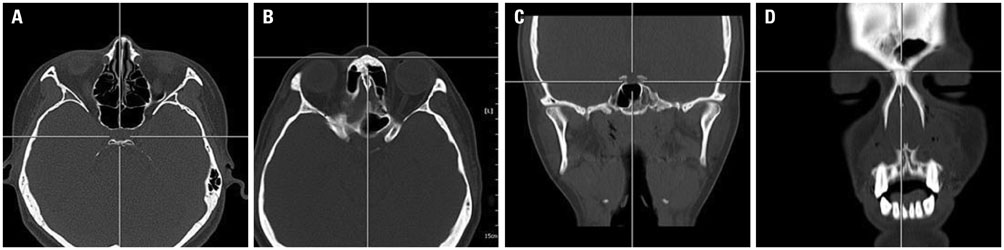
Fig. 2 Three-dimensional (3D) co-ordinates of the sella and nasion in a PACS as the reference line for angular measurement. The 3D co-ordinates of the sella (A) and nasion (B) on axial images were measured using a pixel lens cursor in a PACS report viewer. Each point was verified on reconstructed coronal (C and D) and sagittal images in the PACS report viewer. PACS, picture archiving and communication system.
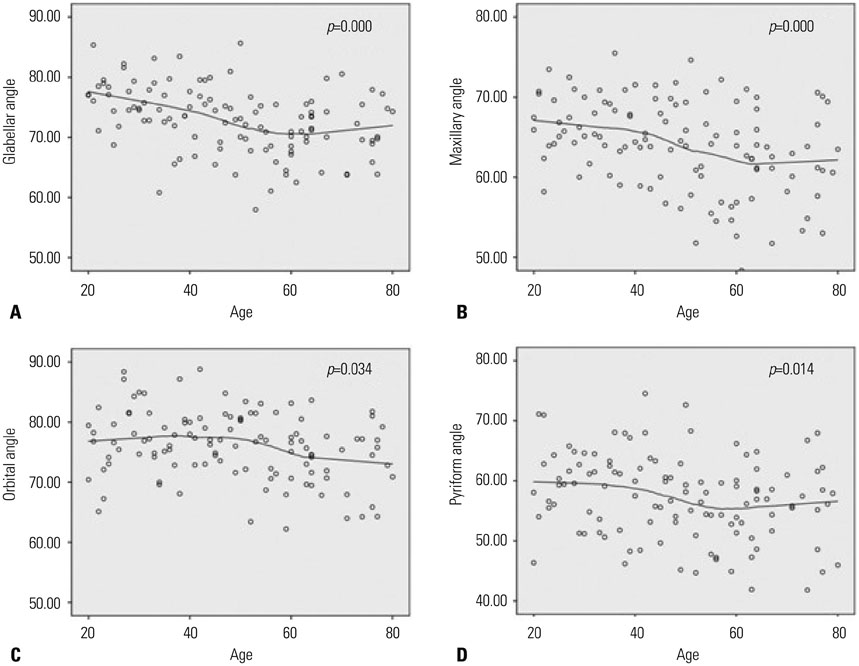
Fig. 3 LOESS regression curves illustrating the trend of changes in glabellar, maxillary, orbital, and pyriform angles based on 115 female data points. Glabellar (A), maxillary (B), orbital (C), and pyriform (D) angles all show statistically significant decreases with aging.

Fig. 4 LOESS regression curves illustrating the trend of changes in glabellar, maxillary, orbital, and pyriform angles based on 108 male data points. Glabellar (A) and maxillary (B) angles show statistically significant decreases with aging, while orbital (C) and pyriform (D) angles do not.

Fig. 5 Mean angular measurements marked on a three-dimensional reconstructed sample image of a female in the middle age group. A prominent zygoma and a wide mandibular angle are seen.
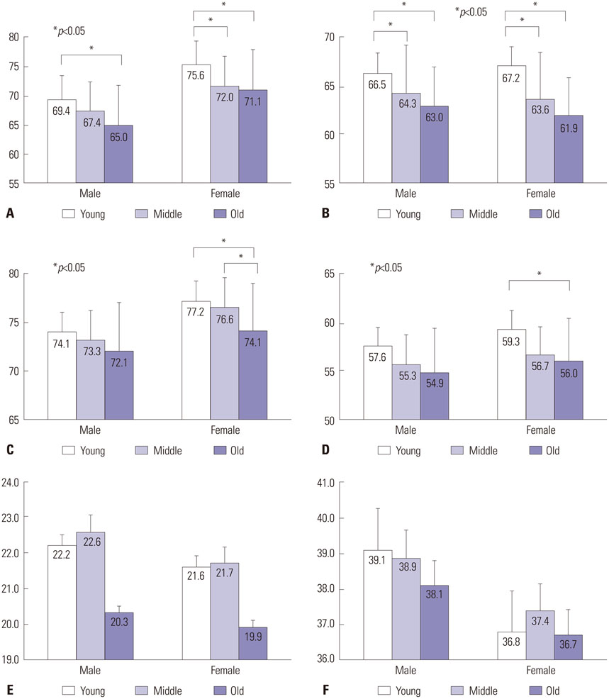
Fig. 6 Column graphs illustrating changes in glabellar angle, maxillary angle, orbital, and pyriform aperture angles, as well as pyriform and orbital widths. Glabellar (A) and maxillary (B) angles show a statistically sinificant decrease with aging in both sexes. Orbital (C) and pyriform (D) angles show a statistically significant decrease with aging in females only. Pyriform (E) and orbital (F) widths do not show statistically significant differences between the age groups.
Cited by 1 articles
-
The Differences in Paranasal Sinus Pneumatization after Adolescence in Korean
Minsu Kang, Ji-Hun Mo, Young-Jun Chung
Korean J Otorhinolaryngol-Head Neck Surg. 2019;62(7):395-403. doi: 10.3342/kjorl-hns.2018.00647.
Reference
-
1. McCurdy JA Jr. Considerations in Asian cosmetic surgery. Facial Plast Surg Clin North Am. 2007; 15:387–397.
Article2. Morris DE, Moaveni Z, Lo LJ. Aesthetic facial skeletal contouring in the Asian patient. Clin Plast Surg. 2007; 34:547–556.
Article3. Wong JK, Larrabee WF Jr. Asian facial plastic surgery. Arch Facial Plast Surg. 2010; 12:217.
Article4. Shirakabe Y, Suzuki Y, Lam SM. A new paradigm for the aging Asian face. Aesthetic Plast Surg. 2003; 27:397–402.
Article5. Lam SM. Aesthetic strategies for the aging Asian face. Facial Plast Surg Clin North Am. 2007; 15:283–291.
Article6. Sykes JM. Management of the aging face in the Asian patient. Facial Plast Surg Clin North Am. 2007; 15:353–360.
Article7. Richard MJ, Morris C, Deen BF, Gray L, Woodward JA. Analysis of the anatomic changes of the aging facial skeleton using computer-assisted tomography. Ophthal Plast Reconstr Surg. 2009; 25:382–386.8. Pessa JE. An algorithm of facial aging: verification of Lambros's theory by three-dimensional stereolithography, with reference to the pathogenesis of midfacial aging, scleral show, and the lateral suborbital trough deformity. Plast Reconstr Surg. 2000; 106:479–488.
Article9. Shaw RB Jr, Kahn DM. Aging of the midface bony elements: a three-dimensional computed tomographic study. Plast Reconstr Surg. 2007; 119:675–681.
Article10. Spiegel M, Lipschutz S, Spellman D. Vector Analysis (Schaum'S Outline). 2nd ed. New York, NY: McGraw-Hill;2009.11. Shaw RB Jr, Katzel EB, Koltz PF, Yaremchuk MJ, Girotto JA, Kahn DM, et al. Aging of the facial skeleton: aesthetic implications and rejuvenation strategies. Plast Reconstr Surg. 2011; 127:374–383.
Article12. Gu Y, McNamara JA Jr, Sigler LM, Baccetti T. Comparison of craniofacial characteristics of typical Chinese and Caucasian young adults. Eur J Orthod. 2011; 33:205–211.
Article13. Yang DB, Chung JY. Infracture technique for reduction malarplasty with a short preauricular incision. Plast Reconstr Surg. 2004; 113:1253–1261.
Article14. Shaw RB Jr, Katzel EB, Koltz PF, Kahn DM, Girotto JA, Langstein HN. Aging of the mandible and its aesthetic implications. Plast Reconstr Surg. 2010; 125:332–342.
Article15. Zadoo VP, Pessa JE. Biological arches and changes to the curvilinear form of the aging maxilla. Plast Reconstr Surg. 2000; 106:460–466.
Article16. Pessa JE, Chen Y. Curve analysis of the aging orbital aperture. Plast Reconstr Surg. 2002; 109:751–755.
Article17. Lee MJ, Popkin BM, Kim S. The unique aspects of the nutrition transition in South Korea: the retention of healthful elements in their traditional diet. Public Health Nutr. 2002; 5:197–203.
Article18. Bogin B, Rios L. Rapid morphological change in living humans: implications for modern human origins. Comp Biochem Physiol A Mol Integr Physiol. 2003; 136:71–84.
Article
- Full Text Links
-
- Actions
-
Cited
- CITED
-
- Close
- Share
-
- Similar articles
-
- Diagnosis of Burst Fracture of the Spine on Plain Radiographs Using the Wintopo(R) Program
- Analysis and Problems of Urolgic Counseling by PC Communication
- A Comparison of Manual and Three-Dimensional Modalities in Predicting Nellix Polymer Volume
- Morphology of the Aging Forehead: A Three-Dimensional Computed Tomographic Study
- The effect of the introduction of Picture Archiving and Communication System on interpretation rate of radiologic examinations
