Clinical and Angiographic Predictors of Microvascular Dysfunction in ST-Segment Elevation Myocardial Infarction
- Affiliations
-
- 1Division of Cardiology, Inha University Hospital, Incheon, Korea. siwoo@inha.ac.kr
- KMID: 2163614
- DOI: http://doi.org/10.3349/ymj.2015.56.5.1235
Abstract
- PURPOSE
We aimed to discover clinical and angiographic predictors of microvascular dysfunction using the index of microcirculatory resistance (IMR) in patients with ST-segment elevation myocardial infarction (STEMI).
MATERIALS AND METHODS
We enrolled 113 patients with STEMI (age, 56+/-11 years; 95 men) who underwent primary percutaneous coronary intervention (PCI). The IMR was measured with a pressure sensor/thermistor-tipped guidewire after primary PCI. The patients were divided into three groups based on IMR values: Low IMR [<18 U (12.9+/-2.6 U), n=38], Mid IMR [18-31 U (23.9+/-4.0 U), n=38], and High IMR [>31 U (48.1+/-17.1 U), n=37].
RESULTS
The age of the Low IMR group was significantly lower than that of the Mid and High IMR groups. The door-to-balloon time was <90 minutes in all patients, and it was not significantly different between groups. Meanwhile, the symptom-onset-to-balloon time was significantly longer in the High IMR group, compared to the Mid and Low IMR groups (p<0.001). In the high IMR group, the culprit lesion was found in a proximal location significantly more often than in a non-proximal location (p=0.008). In multivariate regression analysis, age and symptom-onset-to-balloon time were independent determinants of a high IMR (p=0.013 and p=0.003, respectively).
CONCLUSION
Our data suggest that age and symptom-onset-to-balloon time might be the major predictors of microvascular dysfunction in STEMI patients with a door-to-balloon time of <90 minutes.
Keyword
MeSH Terms
Figure
-
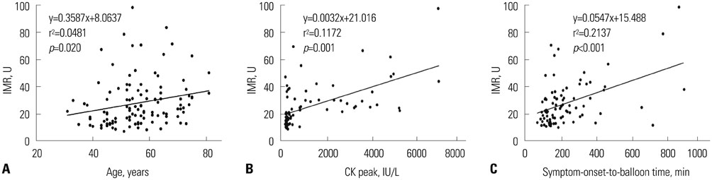
Fig. 1 Relations of age (A), CK peak (B), and symptom-onset-to-balloon time (C) to increasing IMR. Solid lines represent linear regression lines. IMR, index of microcirculatory resistance; CK, creatine kinase.
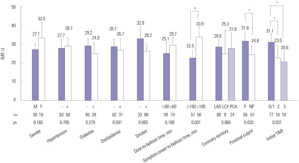
Fig. 2 Comparison of IMR according to clinical and angiographic factors. *The IMR of patients with symptom-onset-to-balloon time of >180 minutes was significantly higher than the IMR of those with a symptom-onset-to-balloon ≤180 minutes, †The IMR was significantly higher in proximal lesion than in non-proximal lesion, ‡The IMR was significantly higher in initial TIMI 0/1 group, as compared initial TIMI 2/3. IMR, index of microcirculatory resistance; LAD, left anterior descending artery; LCX, left circumflex artery; RCA, right coronary artery; P, proximal location of culprit artery; NP, non-proximal location of culprit artery; TIMI, thrombolysis in myocardial infarction.
Cited by 2 articles
-
Therapeutic Hypothermia for Cardioprotection in Acute Myocardial Infarction
In Sook Kang, Ikeno Fumiaki, Wook Bum Pyun
Yonsei Med J. 2016;57(2):291-297. doi: 10.3349/ymj.2016.57.2.291.Gender Differences in Factors Related to Prehospital Delay in Patients with ST-Segment Elevation Myocardial Infarction
Hee-Sook Kim, Kun-Sei Lee, Sang Jun Eun, Si-Wan Choi, Dae Hyeok Kim, Tae-Ho Park, Kyeong Ho Yun, Dong Heon Yang, Seok Jae Hwang, Ki-Soo Park, Rock Bum Kim
Yonsei Med J. 2017;58(4):710-719. doi: 10.3349/ymj.2017.58.4.710.
Reference
-
1. Herrmann J, Kaski JC, Lerman A. Coronary microvascular dysfunction in the clinical setting: from mystery to reality. Eur Heart J. 2012; 33:2771–2782b.
Article2. Fearon WF, Balsam LB, Farouque HM, Caffarelli AD, Robbins RC, Fitzgerald PJ, et al. Novel index for invasively assessing the coronary microcirculation. Circulation. 2003; 107:3129–3132.
Article3. Leung DY, Leung M. Non-invasive/invasive imaging: significance and assessment of coronary microvascular dysfunction. Heart. 2011; 97:587–595.
Article4. Niccoli G, Burzotta F, Galiuto L, Crea F. Myocardial no-reflow in humans. J Am Coll Cardiol. 2009; 54:281–292.
Article5. Gibson CM, Cannon CP, Murphy SA, Ryan KA, Mesley R, Marble SJ, et al. Relationship of TIMI myocardial perfusion grade to mortality after administration of thrombolytic drugs. Circulation. 2000; 101:125–130.
Article6. Wu KC, Zerhouni EA, Judd RM, Lugo-Olivieri CH, Barouch LA, Schulman SP, et al. Prognostic significance of microvascular obstruction by magnetic resonance imaging in patients with acute myocardial infarction. Circulation. 1998; 97:765–772.
Article7. Herzog BA, Husmann L, Valenta I, Gaemperli O, Siegrist PT, Tay FM, et al. Long-term prognostic value of 13N-ammonia myocardial perfusion positron emission tomography added value of coronary flow reserve. J Am Coll Cardiol. 2009; 54:150–156.
Article8. Fearon WF, Shah M, Ng M, Brinton T, Wilson A, Tremmel JA, et al. Predictive value of the index of microcirculatory resistance in patients with ST-segment elevation myocardial infarction. J Am Coll Cardiol. 2008; 51:560–565.
Article9. McGeoch R, Watkins S, Berry C, Steedman T, Davie A, Byrne J, et al. The index of microcirculatory resistance measured acutely predicts the extent and severity of myocardial infarction in patients with ST-segment elevation myocardial infarction. JACC Cardiovasc Interv. 2010; 3:715–722.10. Yoo SH, Yoo TK, Lim HS, Kim MY, Koh JH. Index of microcirculatory resistance as predictor for microvascular functional recovery in patients with anterior myocardial infarction. J Korean Med Sci. 2012; 27:1044–1050.
Article11. Fearon WF, Low AF, Yong AS, McGeoch R, Berry C, Shah MG, et al. Prognostic value of the Index of Microcirculatory Resistance measured after primary percutaneous coronary intervention. Circulation. 2013; 127:2436–2441.
Article12. O'Gara PT, Kushner FG, Ascheim DD, Casey DE Jr, Chung MK, de Lemos JA, et al. 2013 ACCF/AHA guideline for the management of ST-elevation myocardial infarction: a report of the American College of Cardiology Foundation/American Heart Association Task Force on Practice Guidelines. Circulation. 2013; 127:e362–e425.13. Wilson RF, Wyche K, Christensen BV, Zimmer S, Laxson DD. Effects of adenosine on human coronary arterial circulation. Circulation. 1990; 82:1595–1606.
Article14. Pijls NH, De Bruyne B, Smith L, Aarnoudse W, Barbato E, Bartunek J, et al. Coronary thermodilution to assess flow reserve: validation in humans. Circulation. 2002; 105:2482–2486.15. Pijls NH, De Bruyne B, Peels K, Van Der Voort PH, Bonnier HJ, Bartunek J, Koolen JJ, et al. Measurement of fractional flow reserve to assess the functional severity of coronary-artery stenoses. N Engl J Med. 1996; 334:1703–1708.
Article16. De Bruyne B, Pijls NH, Smith L, Wievegg M, Heyndrickx GR. Coronary thermodilution to assess flow reserve: experimental validation. Circulation. 2001; 104:2003–2006.17. Lang RM, Bierig M, Devereux RB, Flachskampf FA, Foster E, Pellikka PA, et al. Recommendations for chamber quantification: a report from the American Society of Echocardiography’s Guidelines and Standards Committee and the Chamber Quantification Writing Group, developed in conjunction with the European Association of Echocardiography, a branch of the European Society of Cardiology. J Am Soc Echocardiogr. 2005; 18:1440–1463.
Article18. O'Rourke MF. Arterial aging: pathophysiological principles. Vasc Med. 2007; 12:329–341.19. Rizzoni D, Porteri E, Boari GE, De Ciuceis C, Sleiman I, Muiesan ML, et al. Prognostic significance of small-artery structure in hypertension. Circulation. 2003; 108:2230–2235.
Article20. Safar ME. Peripheral pulse pressure, large arteries, and microvessels. Hypertension. 2004; 44:121–122.21. Potenza MA, Gagliardi S, Nacci C, Carratu' MR, Montagnani M. Endothelial dysfunction in diabetes: from mechanisms to therapeutic targets. Curr Med Chem. 2009; 16:94–112.
Article22. Marciano C, Galderisi M, Gargiulo P, Acampa W, D'Amore C, Esposito R, et al. Effects of type 2 diabetes mellitus on coronary microvascular function and myocardial perfusion in patients without obstructive coronary artery disease. Eur J Nucl Med Mol Imaging. 2012; 39:1199–1206.
Article23. Rathore SS, Curtis JP, Chen J, Wang Y, Nallamothu BK, Epstein AJ, et al. Association of door-to-balloon time and mortality in patients admitted to hospital with ST elevation myocardial infarction: national cohort study. BMJ. 2009; 338:b1807.
Article24. McNamara RL, Wang Y, Herrin J, Curtis JP, Bradley EH, Magid DJ, et al. Effect of door-to-balloon time on mortality in patients with ST-segment elevation myocardial infarction. J Am Coll Cardiol. 2006; 47:2180–2186.
Article25. De Luca G, Suryapranata H, Ottervanger JP, Antman EM. Time delay to treatment and mortality in primary angioplasty for acute myocardial infarction: every minute of delay counts. Circulation. 2004; 109:1223–1225.
Article26. O'Gara PT, Kushner FG, Ascheim DD, Casey DE Jr, Chung MK, de Lemos JA, et al. 2013 ACCF/AHA guideline for the management of ST-elevation myocardial infarction: executive summary: a report of the American College of Cardiology Foundation/American Heart Association Task Force on Practice Guidelines. Circulation. 2013; 127:529–555.27. Menees DS, Peterson ED, Wang Y, Curtis JP, Messenger JC, Rumsfeld JS, et al. Door-to-balloon time and mortality among patients undergoing primary PCI. N Engl J Med. 2013; 369:901–909.
Article28. Gibson CM, Schömig A. Coronary and myocardial angiography: angiographic assessment of both epicardial and myocardial perfusion. Circulation. 2004; 109:3096–3105.29. Angeja BG, Gunda M, Murphy SA, Sobel BE, Rundle AC, Syed M, et al. TIMI myocardial perfusion grade and ST segment resolution: association with infarct size as assessed by single photon emission computed tomography imaging. Circulation. 2002; 105:282–285.
Article30. Lim HS, Yoon MH, Tahk SJ, Yang HM, Choi BJ, Choi SY, et al. Usefulness of the index of microcirculatory resistance for invasively assessing myocardial viability immediately after primary angioplasty for anterior myocardial infarction. Eur Heart J. 2009; 30:2854–2860.
Article31. Byrne RA, Ndrepepa G, Braun S, Tiroch K, Mehilli J, Schulz S, et al. Peak cardiac troponin-T level, scintigraphic myocardial infarct size and one-year prognosis in patients undergoing primary percutaneous coronary intervention for acute myocardial infarction. Am J Cardiol. 2010; 106:1212–1217.
Article32. Chin CT, Wang TY, Li S, Wiviott SD, deLemos JA, Kontos MC, et al. Comparison of the prognostic value of peak creatine kinase-MB and troponin levels among patients with acute myocardial infarction: a report from the Acute Coronary Treatment and Intervention Outcomes Network Registry-get with the guidelines. Clin Cardiol. 2012; 35:424–429.
Article33. Tamita K, Akasaka T, Takagi T, Yamamuro A, Yamabe K, Katayama M, et al. Effects of microvascular dysfunction on myocardial fractional flow reserve after percutaneous coronary intervention in patients with acute myocardial infarction. Catheter Cardiovasc Interv. 2002; 57:452–459.
Article34. Ntalianis A, Sels JW, Davidavicius G, Tanaka N, Muller O, Trana C, et al. Fractional flow reserve for the assessment of nonculprit coronary artery stenoses in patients with acute myocardial infarction. JACC Cardiovasc Interv. 2010; 3:1274–1281.
Article35. Gibson CM, Pinto D. Fractional flow reserve: a new set of lenses for the occulostenotic reflex? JACC Cardiovasc Interv. 2010; 3:1282–1283.36. Murai T, Lee T, Yonetsu T, Iwai T, Takagi T, Hishikari K, et al. Variability of microcirculatory resistance index and its relationship with fractional flow reserve in patients with intermediate coronary artery lesions. Circ J. 2013; 77:1769–1776.
Article37. Harjai KJ, Mehta RH, Stone GW, Boura JA, Grines L, Brodie BR, et al. Does proximal location of culprit lesion confer worse prognosis in patients undergoing primary percutaneous coronary intervention for ST elevation myocardial infarction? J Interv Cardiol. 2006; 19:285–294.
Article38. Vlaar PJ, Svilaas T, van der Horst IC, Diercks GF, Fokkema ML, de Smet BJ, et al. Cardiac death and reinfarction after 1 year in the Thrombus Aspiration during Percutaneous coronary intervention in Acute myocardial infarction Study (TAPAS): a 1-year follow-up study. Lancet. 2008; 371:1915–1920.
Article39. Fröbert O, Lagerqvist B, Olivecrona GK, Omerovic E, Gudnason T, Maeng M, et al. Thrombus aspiration during ST-segment elevation myocardial infarction. N Engl J Med. 2013; 369:1587–1597.
Article40. Woo SI, Park SD, Kim DH, Kwan J, Shin SH, Park KS, et al. Thrombus aspiration during primary percutaneous coronary intervention for preserving the index of microcirculatory resistance: a randomised study. EuroIntervention. 2014; 9:1057–1062.
Article41. Sethi A, Bajaj A, Bahekar A, Bhuriya R, Singh M, Ahmed A, et al. Glycoprotein IIb/IIIa inhibitors with or without thienopyridine pretreatment improve outcomes after primary percutaneous coronary intervention in high-risk patients with ST elevation myocardial infarction--a meta-regression of randomized controlled trials. Catheter Cardiovasc Interv. 2013; 82:171–181.
Article
- Full Text Links
-
- Actions
-
Cited
- CITED
-
- Close
- Share
-
- Similar articles
-
- Erratum: Etiologies and Predictors of False-Positive Diagnosis of ST-Segment Elevation Myocardial Infarction
- Effect of Aspiration Thrombectomy on Microvascular Dysfunction in ST-Segment Elevation Myocardial Infarction With an Elevated Neutrophil Count
- ST segment
- A Case of ST-Segment Elevation in a Patient with Subarachnoid Hemorrhage
- Impact of ST segment deviation in patients with acute myocardial infarction and left ventricular systolic dysfunction
