The utility of the 3D imaging software in the macroscopic rendering of complex gynecologic specimens
- Affiliations
-
- 1Department of Diagnostic and Clinical Medicine, University of Modena and Reggio Emilia, Modena, Italy. emailmedical@gmail.com
- 2Department of General Surgery and Surgical Specialties, University of Modena and Reggio Emilia, Modena, Italy.
- 3Provincial Health Care Services, Institute of Pathology, Santa Maria del Carmine Hospital, Rovereto, Italy.
- 4Department of Radiology, University of Perugia, Perugia, Italy.
- KMID: 2160801
- DOI: http://doi.org/10.3802/jgo.2015.26.2.168
Abstract
- No abstract available.
MeSH Terms
-
Abdomen/pathology/surgery
Adult
Endometrial Neoplasms/complications/*pathology/radiography/surgery
Endometriosis/complications/*pathology/radiography/surgery
Female
Humans
Image Enhancement/*methods
Imaging, Three-Dimensional/*methods
Pelvis/pathology/radiography/surgery
Radiography, Abdominal
Sarcoma, Endometrial Stromal/complications/*pathology/radiography/surgery
*Software
Specimen Handling
Figure
-
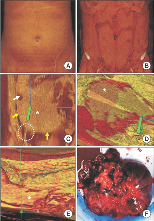
Fig. 1 (A) A longitudinal digitalized image of the abdomen, centered on the umbilicus, is seen without application of subtraction filters. (B) The dermal subtraction filter allows to remove the skin surface, which does not show pathological alterations. (C) The superficial muscle subtraction filter is able to subtract the superficial muscle layer and the sarcoma appears as a right-sided abdominal bulky mass (asterisk). The neoplasia shows a pseudo-capsule (yellow arrows), an area of colliquation (white arrow) and take its origin from the right inguinal ligament (white circle). The green pointer of spatial orientation is also reported. (D) The deep muscle subtraction filter points out the infiltration of the surrounding deep tissues, which appears as a color distortion (asterisk). (E) On a transversal section, the visceral subtraction filter displays the presence of a massive front of neoplastic infiltration towards the intestine (asterisk). (F) The gross appearance confirms what has been previously observed by the digitalized images.
Reference
-
1. Zeller JL. New 3D imaging software opens new vistas. JAMA. 2006; 296:2908–2913.2. Bergamini A, Almirante G, Taccagni G, Mangili G, Vigano P, Candiani M. Endometriosis-associated tumor at the inguinal site: report of a case diagnosed during pregnancy and literature review. J Obstet Gynaecol Res. 2014; 40:1132–1136.3. Kim JY, Hong SY, Sung HJ, Oh HK, Koh SB. A case of multiple metastatic low-grade endometrial stromal sarcoma arising from an ovarian endometriotic lesion. J Gynecol Oncol. 2009; 20:122–125.4. Alcazar JL, Guerriero S, Ajossa S, Parodo G, Piras B, Peiretti M, et al. Extragenital endometrial stromal sarcoma arising in endometriosis. Gynecol Obstet Invest. 2012; 73:265–271.5. Sato K, Ueda Y, Sugaya J, Ozaki M, Hisaoka M, Katsuda S. Extrauterine endometrial stromal sarcoma with JAZF1/JJAZ1 fusion confirmed by RT-PCR and interphase FISH presenting as an inguinal tumor. Virchows Arch. 2007; 450:349–353.6. Sinha R, Sundaram M. Endometrial stromal sarcoma from endometriosis. J Minim Invasive Gynecol. 2010; 17:541–542.7. Usta TA, Sonmez SE, Oztarhan A, Karacan T. Endometrial stromal sarcoma in the abdominal wall arising from scar endometriosis. J Obstet Gynaecol. 2014; 34:541–542.
- Full Text Links
-
- Actions
-
Cited
- CITED
-
- Close
- Share
-
- Similar articles
-
- Clinical Research through Computational Anatomy and Virtual Fixation
- Three-Dimensional Images and Software for Studying Anatomical Structures in MRIs
- Evaluation of accuracy of 3D reconstruction images using multi-detector CT and cone-beam CT
- Usefulness of PC Based 3D Volume Rendering Technique in the Evaluation of Suspected Aneurysm on Brain MRA
- Diagnostic Accuracy of the Volume Rendering Images of Multi-Detector CT for the Detection of Lumbar Transverse Process Fractures
