Imaging Sci Dent.
2012 Mar;42(1):25-33. 10.5624/isd.2012.42.1.25.
Evaluation of accuracy of 3D reconstruction images using multi-detector CT and cone-beam CT
- Affiliations
-
- 1Department of Orthodontics, Hangang Sacred Heart Hospital, Graduate School of Clinical Dentistry, Hallym University, Seoul, Korea.
- 2Department of Oral and Maxillofacial Radiology and Dental Research Institute, School of Dentistry, Seoul National University, Seoul, Korea. future3@snu.ac.kr
- 3Department of Oral and Maxillofacial Radiology, BK21 Craniomaxillofacial Life Science, and Dental Research Institute, School of Dentistry, Seoul National University, Seoul, Korea.
- KMID: 1974404
- DOI: http://doi.org/10.5624/isd.2012.42.1.25
Abstract
- PURPOSE
This study was performed to determine the accuracy of linear measurements on three-dimensional (3D) images using multi-detector computed tomography (MDCT) and cone-beam computed tomography (CBCT).
MATERIALS AND METHODS
MDCT and CBCT were performed using 24 dry skulls. Twenty-one measurements were taken on the dry skulls using digital caliper. Both types of CT data were imported into OnDemand software and identification of landmarks on the 3D surface rendering images and calculation of linear measurements were performed. Reproducibility of the measurements was assessed using repeated measures ANOVA and ICC, and the measurements were statistically compared using a Student t-test.
RESULTS
All assessments under the direct measurement and image-based measurements on the 3D CT surface rendering images using MDCT and CBCT showed no statistically difference under the ICC examination. The measurements showed no differences between the direct measurements of dry skull and the image-based measurements on the 3D CT surface rendering images (P>.05).
CONCLUSION
Three-dimensional reconstructed surface rendering images using MDCT and CBCT would be appropriate for 3D measurements.
MeSH Terms
Figure
-
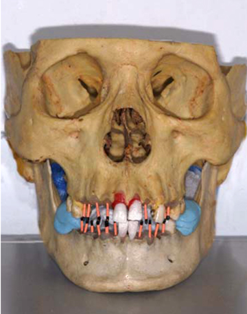
Fig. 1 An example of dry skull specimen with mandible fixed to the cranium.

Fig. 2 An example of MDCT 3D image (left) and CBCT 3D image (right) on OnDemend™ (Cybermed, Seoul, Korea) software.
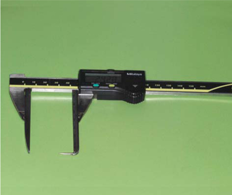
Fig. 3 A modified digital caliper to measure distance between landmarks located in concave surface.
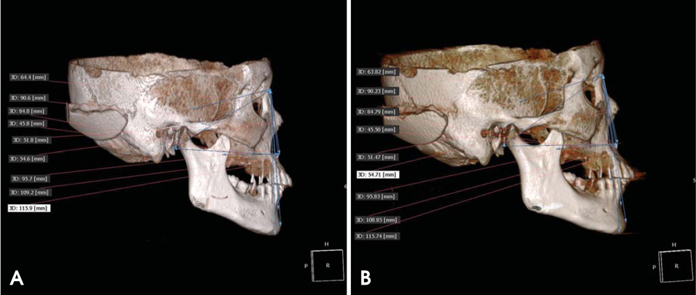
Fig. 4 An example of measurement between mid-sagittal landmarks using OnDemand. A. MDCT. B. CBCT.
Cited by 1 articles
-
Linear accuracy of cone-beam computed tomography and a 3-dimensional facial scanning system: An anthropomorphic phantom study
Song Hee Oh, Ju Hee Kang, Yu-Kyeong Seo, Sae Rom Lee, Hwa-Young Choi, Yong-Suk Choi, Eui-Hwan Hwang
Imaging Sci Dent. 2018;48(2):111-119. doi: 10.5624/isd.2018.48.2.111.
Reference
-
1. Mozzo P, Procacci C, Tacconi A, Martini PT, Andreis IA. A new volumetric CT machine for dental imaging based on the cone-beam technique: preliminary results. Eur Radiol. 1998. 8:1558–1564.
Article2. Sukovic P. Cone beam computed tomography in craniofacial imaging. Orthod Craniofac Res. 2003. 6:suppl 1. 31–36.
Article3. Ludlow JB, Davies-Ludlow LE, Brooks SL, Howerton WB. Dosimetry of 3 CBCT devices for oral and maxillofacial radiology: CB Mercuray, NewTom 3G and i-CAT. Dentomaxillofac Radiol. 2006. 35:219–226.
Article4. Maki K, Inou N, Takanishi A, Miller AJ. Computer-assisted simulations in orthodontic diagnosis and the application of a new cone beam X-ray computed tomography. Orthod Craniofac Res. 2003. 6:suppl 1. 95–101.
Article5. Aboudara CA, Hatcher D, Nielsen IL, Miller A. A three-dimensional evaluation of the upper airway in adolescents. Orthod Craniofac Res. 2003. 6:suppl 1. 173–175.
Article6. Cevidanes LH, Styner MA, Proffit WR. Image analysis and superimposition of 3-dimensional cone-beam computed tomography models. Am J Orthod Dentofacial Orthop. 2006. 129:611–618.
Article7. Kau CH, Richmond S, Palomo JM, Hans MG. Three-dimensional cone beam computerized tomography in orthodontics. J Orthod. 2005. 32:282–293.8. Park SH, Yu HS, Kim KD, Lee KJ, Baik HS. A proposal for a new analysis of craniofacial morphology by 3-dimensional computed tomography. Am J Orthod Dentofacial Orthop. 2006. 129:600.e23–600.e34.
Article9. Maeda M, Katsumata A, Ariji Y, Muramatsu A, Yoshida K, Goto S, et al. 3D-CT evaluation of facial asymmetry in patients with maxillofacial deformities. Oral Surg Oral Med Oral Pathol Oral Radiol Endod. 2006. 102:382–390.
Article10. Olszewski R, Cosnard G, Macq B, Mahy P, Reychler H. 3D CT-based cephalometric analysis: 3D cephalometric theoretical concept and software. Neuroradiology. 2006. 48:853–862.
Article11. Hwang HS, Hwang CH, Lee KH, Kang BC. Maxillofacial 3-dimensional image analysis for the diagnosis of facial asymmetry. Am J Orthod Dentofacial Orthop. 2006. 130:779–785.
Article12. Lagravère MO, Hansen L, Harzer W, Major PW. Plane orientation for standardization in 3-dimensional cephalometric analysis with computerized tomography imaging. Am J Orthod Dentofacial Orthop. 2006. 129:601–604.
Article13. Swennen GR, Schutyser F, Barth EL, De Groeve P, De Mey A. A new method of 3-D cephalometry Part I: The anatomic Cartesian 3-D reference system. J Craniofac Surg. 2006. 17:314–325.14. Olszewski R, Zech F, Cosnard G, Nicolas V, Macq B, Reychler H. Three-dimensional computed tomography cephalometric craniofacial analysis: experimental validation in vitro. Int J Oral Maxillofac Surg. 2007. 36:828–833.
Article15. Yoon SJ, Lim HJ, Kang BC, Hwang HS. Three dimensional CT analysis of facial asymmetry. Korean J Oral Maxillofac Radiol. 2007. 37:45–51.16. Xia J, Samman N, Yeung RW, Shen SG, Wang D, Ip HH, et al. Three-dimensional virtual reality surgical planning and simulation workbench for orthognathic surgery. Int J Adult Orthodon Orthognath Surg. 2000. 15:265–282.17. Westermark A, Zachow S, Eppley BL. Three-dimensional osteotomy planning in maxillofacial surgery including soft tissue prediction. J Craniofac Surg. 2005. 16:100–104.
Article18. Hildebolt CF, Vannier MW, Knapp RH. Validation study of skull three-dimensional computerized tomography measurements. Am J Phys Anthropol. 1990. 82:283–294.
Article19. Kragskov J, Bosch C, Gyldensted C, Sindet-Pedersen S. Comparison of the reliability of craniofacial anatomic landmarks based on cephalometric radiographs and three-dimensional CT scans. Cleft Palate Craniofac J. 1997. 34:111–116.
Article20. Nagashima M, Inoue K, Sasaki T, Miyasaka K, Matsumura G, Kodama G. Three-dimensional imaging and osteometry of adult human skulls using helical computed tomography. Surg Radiol Anat. 1998. 20:291–297.
Article21. Cavalcanti MG, Vannier MW. Quantitative analysis of spiral computed tomography for craniofacial clinical applications. Dentomaxillofac Radiol. 1998. 27:344–350.
Article22. Cavalcanti MG, Haller JW, Vannier MW. Three-dimensional computed tomography landmark measurement in craniofacial surgical planning: experimental validation in vitro. J Oral Maxillofac Surg. 1999. 57:690–694.
Article23. Jung H, Kim HJ, Kim DO, Hong SI, Jeong HK, Kim KD, et al. Quantitative analysis of three-dimensional rendered imaging of the human skull acquired from multi-detector row computed tomography. J Digit Imaging. 2002. 15:232–239.
Article24. Kim DO, Kim HJ, Jung H, Jeong HK, Hong SI, Kim KD. Quantitative evaluation of acquisition parameters in three-dimensional imaging with multidetector computed tomography using human skull phantom. J Digit Imaging. 2002. 15:suppl 1. 254–257.
Article25. Jeon KJ, Park H, Lee HC, Kim KD, Park CS. Reproducibilities of cephalometric measurements of three-dimensional CT images reconstructed in the personal computer. Korean J Oral Maxillofac Radiol. 2003. 33:171–178.26. Williams FL, Richtsmeier JT. Comparison of mandibular landmarks from computed tomography and 3D digitizer data. Clin Anat. 2003. 16:494–500.
Article27. Cavalcanti MG, Rocha SS, Vannier MW. Craniofacial measurements based on 3D-CT volume rendering: implications for clinical applications. Dentomaxillofac Radiol. 2004. 33:170–176.
Article28. Park JW, Kim NK, Chang YI. Formulation of a reference coordinate system of three-dimensional (3D) head & neck images: Part I. Reproducibility of 3D cephalometric landmarks. Korean J Orthod. 2005. 35:388–397.29. Olszewski R, Reychler H, Cosnard G, Denis JM, Vynckier S, Zech F. Accuracy of three-dimensional (3D) craniofacial cephalometric landmarks on a low-dose 3D computed tomograph. Dentomaxillofac Radiol. 2008. 37:261–267.
Article30. Lopes PM, Moreira CR, Perrella A, Antunes JL, Cavalcanti MG. 3-D volume rendering maxillofacial analysis of angular measurements by multislice CT. Oral Surg Oral Med Oral Pathol Oral Radiol Endod. 2008. 105:224–230.
Article31. Stratemann SA, Huang JC, Maki K, Miller AJ, Hatcher DC. Comparison of cone beam computed tomography imaging with physical measures. Dentomaxillofac Radiol. 2008. 37:80–93.
Article32. Periago DR, Scarfe WC, Moshiri M, Scheetz JP, Silveira AM, Farman AG. Linear accuracy and reliability of cone beam CT derived 3-dimensional images constructed using an orthodontic volumetric rendering program. Angle Orthod. 2008. 78:387–395.
Article33. Brown AA, Scarfe WC, Scheetz JP, Silveira AM, Farman AG. Linear accuracy of cone beam CT derived 3D images. Angle Orthod. 2009. 79:150–157.
Article34. Kumar V, Ludlow JB, Mol A, Cevidanes L. Comparison of conventional and cone beam CT synthesized cephalograms. Dentomaxillofac Radiol. 2007. 36:263–269.
Article35. Olszewski R, Tanesy O, Cosnard G, Zech F, Reychler H. Reproducibility of osseous landmarks used for computed tomography based three-dimensional cephalometric analyses. J Craniomaxillofac Surg. 2010. 38:214–221.
Article36. Baumrind S, Frantz RC. The reliability of head film measurements. 1. Landmark identification. Am J Orthod. 1971. 60:111–127.37. Chien PC, Parks ET, Eraso F, Hartsfield JK, Roberts WE, Ofner S. Comparison of reliability in anatomical landmark identi-fication using two-dimensional digital cephalometrics and three-dimensional cone beam computed tomography in vivo. Dentomaxillofac Radiol. 2009. 38:262–273.38. Jeong HG, Kim KD, Park H, Kim DO, Jeong H, Kim HJ, et al. Three-dimensional image analysis of the skull using variable CT scanning protocols-effect of slice thickness on measurement in the three-dimensional CT images. Korean J Oral Maxillofac Radiol. 2004. 34:151–157.39. Swennen GR, Schutyser F. Three-dimensional cephalometry: spiral multi-slice vs cone-beam computed tomography. Am J Orthod Dentofacial Orthop. 2006. 130:410–416.
Article40. Richtsmeier JT, Paik CH, Elfert PC, Cole TM 3rd, Dahlman HR. Precision, repeatability, and validation of the localization of cranial landmarks using computed tomography scans. Cleft Palate Craniofac J. 1995. 32:217–227.
Article41. Fox LA, Vannier MW, West OC, Wilson AJ, Baran GA, Pilgram TK. Diagnostic performance of CT, MPR, 3DCT imaging in maxillofacial trauma. Comput Med Imaging Graph. 1995. 19:385–395.42. Laine FJ, Conway WF, Laskin DM. Radiology of maxillofacial trauma. Curr Probl Diagn Radiol. 1993. 22:145–188.
Article
- Full Text Links
-
- Actions
-
Cited
- CITED
-
- Close
- Share
-
- Similar articles
-
- Multi-Detector Row CT of the Central Airway Disease
- Comparison of CT numbers between cone-beam CT and multi-detector CT
- Diagnostic Accuracy of the Volume Rendering Images of Multi-Detector CT for the Detection of Lumbar Transverse Process Fractures
- Three dimensional evaluation of impacted mesiodens using dental cone beam CT
- A Study on the Availability of the On-Board Imager (OBI) and Cone-Beam CT (CBCT) in the Verification of Patient Set-up
