Three Dimensional Quantitative Coronary Angiography Can Detect Reliably Ischemic Coronary Lesions Based on Fractional Flow Reserve
- Affiliations
-
- 1Devision of Cardiology, Department of Internal Medicine, Boramae Medical Center, Seoul National University, College of Medicine, Seoul, Korea.
- 2Department of Cardiology, Ajou University School of Medicine, Suwon, Korea.
- 3Division of Cardiology, Department of Internal Medicine, School of Medicine, Dankook University, Cheonan, Korea.
- 4Department of Cardiovascular Medicine, Wakayama Medical University, Wakayama, Japan.
- 5Division of Cardiovascular Disease, Mayo Clinic, Rochester, MN, USA. Lerman.Amir@mayo.edu
- KMID: 2160599
- DOI: http://doi.org/10.3346/jkms.2015.30.6.716
Abstract
- Conventional coronary angiography (CAG) has limitations in evaluating lesions producing ischemia. Three dimensional quantitative coronary angiography (3D-QCA) shows reconstructed images of CAG using computer based algorithm, the Cardio-op B system (Paieon Medical, Rosh Ha'ayin, Israel). The aim of this study was to evaluate whether 3D-QCA can reliably predict ischemia assessed by myocardial fractional flow reserve (FFR) < 0.80. 3D-QCA images were reconstructed from CAG which also were evaluated with FFR to assess ischemia. Minimal luminal diameter (MLD), percent diameter stenosis (%DS), minimal luminal area (MLA), and percent area stenosis (%AS) were obtained. The results of 3D-QCA and FFR were compared. A total of 266 patients was enrolled for the present study. FFR for all lesions ranged from 0.57 to 1.00 (0.85 +/- 0.09). Measurement of MLD, %DS, MLA, and %AS all were significantly correlated with FFR (r = 0.569, 0609, 0.569, 0.670, respectively, all P < 0.001). In lesions with MLA < 4.0 mm2, %AS of more than 65.5% had a 80% sensitivity and a 83% specificity to predict FFR < 0.80 (area under curve, AUC was 0.878). 3D-QCA can reliably predict coronary lesions producing ischemia and may be used to guide therapeutic approach for coronary artery disease.
MeSH Terms
-
Aged
Coronary Angiography/*methods
Coronary Circulation
Coronary Stenosis/etiology/*physiopathology/*radiography
Female
*Fractional Flow Reserve, Myocardial
Humans
Imaging, Three-Dimensional/*methods
Male
Myocardial Ischemia/complications/physiopathology/*radiography
Radiographic Image Enhancement/methods
Radiographic Image Interpretation, Computer-Assisted/methods
Reproducibility of Results
Sensitivity and Specificity
Figure
-

Fig. 1 Reconstruction of three dimensional images. (A) Calibration is done with catheter seen on the same image. (B, C) Clicking the narrowest point, proximal and distal reference segment make the lesion lineated automatically. It should be done on two images at least 30 degree apart from each other. (D-F), After finishing (A-C), computer based algorithm is activated and shows three dimensional images and quantitative data.
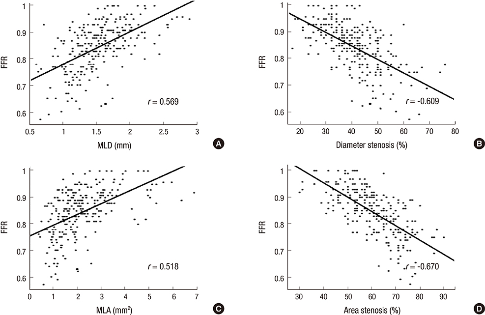
Fig. 2 Bivariate linear correlation of 3D-QCA variables with FFR. (A) MLD, (B) %DS, (C) MLA, and (D) %AS. 3D-QCA, three-dimensional quantitative coronary angiography; FFR, fractional flow reserve; MLD, minimal luminal diameter; %DS, percent diameter stenosis; MLA, minimal luminal area; %AS, percent area stenosis; r, Pearson's correlation coefficient.
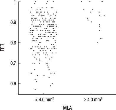
Fig. 3 Difference in FFR according to MLA divided by 4.0 mm2 on 3D-QCA. 3D-QCA, three-dimensional quantitative coronary angiography; MLA, minimal luminal area; FFR, fractional flow reserve.
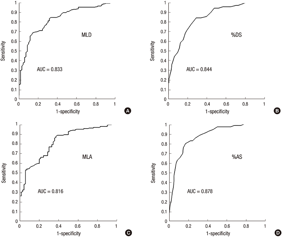
Fig. 4 Receiver operating characteristic curve of 3D-QCA variables in lesions with MLA < 4.0 mm2 for FFR < 0.80. (A) Minimal luminal diameter. (B) percent area stenosis. (C) minimal luminal area. (D) percent area stenosis. 3D-QCA, three-dimensional quantitative coronary angiography; FFR, fractional flow reserve; MLD, minimal luminal diameter; %DS, percent diameter stenosis; MLA, minimal luminal area; %AS, percent area stenosis; AUC, area under curve.
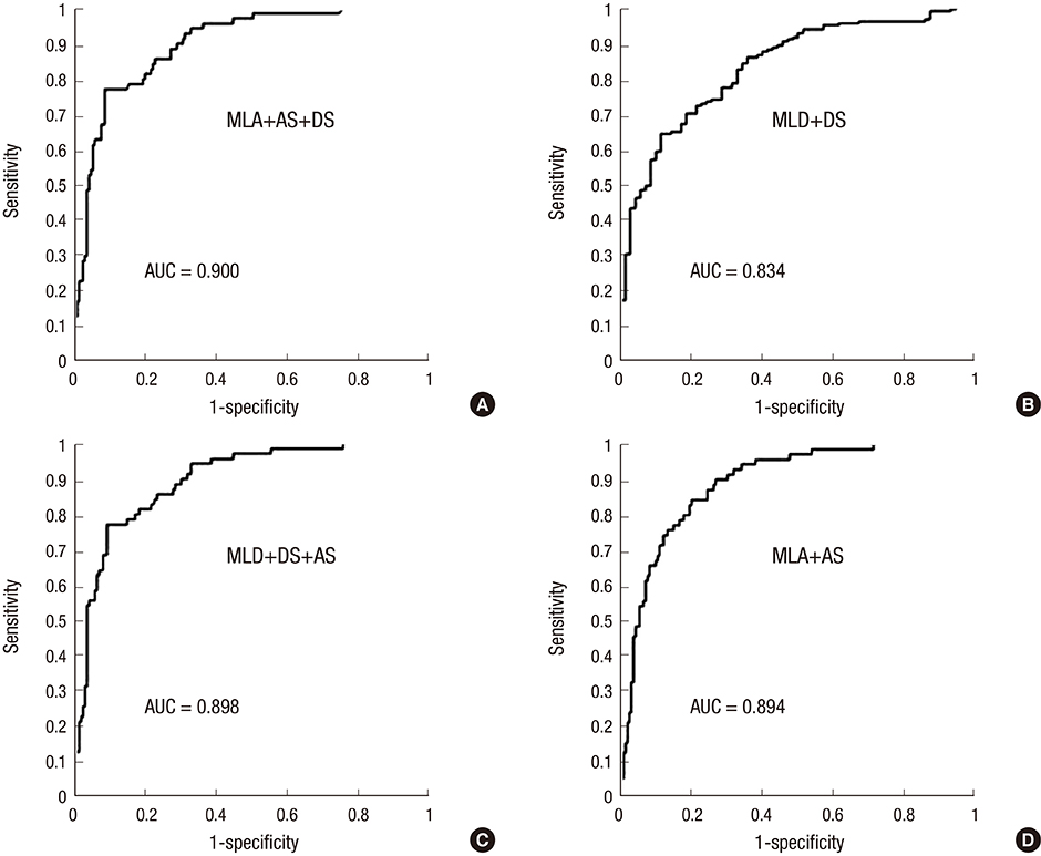
Fig. 5 Receiver operating characteristic curve of combined 3D-QCA variables in lesions with MLA < 4.0 mm2 for FFR < 0.80. (A) MLA plus %AS plus %DS. (B) MLD plus %DS. (C) MLD plus %DS plus %AS. (D) MLA plus %AS. 3D-QCA, three-dimensional quantitative coronary angiography; FFR, fractional flow reserve; MLD, minimal luminal diameter; %DS, percent diameter stenosis; MLA, minimal luminal area; %AS, percent area stenosis; AUC, area under curve.
Reference
-
1. Gottsauner-Wolf M, Sochor H, Moertl D, Gwechenberger M, Stockenhuber F, Probst P. Assessing coronary stenosis. Quantitative coronary angiography versus visual estimation from cine-film or pharmacological stress perfusion images. Eur Heart J. 1996; 17:1167–1174.2. Topol EJ, Nissen SE. Our preoccupation with coronary luminology. The dissociation between clinical and angiographic findings in ischemic heart disease. Circulation. 1995; 92:2333–2342.3. Pijls NH, van Schaardenburgh P, Manoharan G, Boersma E, Bech JW, van't Veer M, Bär F, Hoorntje J, Koolen J, Wijns W, et al. Percutaneous coronary intervention of functionally nonsignificant stenosis: 5-year follow-up of the DEFER Study. J Am Coll Cardiol. 2007; 49:2105–2111.4. Pijls NH, De Bruyne B, Peels K, Van Der Voort PH, Bonnier HJ, Bartunek JKJJ, Koolen JJ. Measurement of fractional flow reserve to assess the functional severity of coronary-artery stenoses. N Engl J Med. 1996; 334:1703–1708.5. Tonino PA, De Bruyne B, Pijls NH, Siebert U, Ikeno F, van't Veer M, Klauss V, Manoharan G, Engstrøm T, Oldroyd KG, et al. FAME Study Investigators. Fractional flow reserve versus angiography for guiding percutaneous coronary intervention. N Engl J Med. 2009; 360:213–224.6. Hamilos M, Muller O, Cuisset T, Ntalianis A, Chlouverakis G, Sarno G, Nelis O, Bartunek J, Vanderheyden M, Wyffels E, et al. Long-term clinical outcome after fractional flow reserve-guided treatment in patients with angiographically equivocal left main coronary artery stenosis. Circulation. 2009; 120:1505–1512.7. Tonino PA, Fearon WF, De Bruyne B, Oldroyd KG, Leesar MA, Ver Lee PN, Maccarthy PA, Van't Veer M, Pijls NH. Angiographic versus functional severity of coronary artery stenoses in the FAME study fractional flow reserve versus angiography in multivessel evaluation. J Am Coll Cardiol. 2010; 55:2816–2821.8. Agostoni P, Biondi-Zoccai G, Van Langenhove G, Cornelis K, Vermeersch P, Convens C, Vassanelli C, Van Den Heuvel P, Van Den Branden F, Verheye S. Comparison of assessment of native coronary arteries by standard versus three-dimensional coronary angiography. Am J Cardiol. 2008; 102:272–279.9. Dvir D, Marom H, Guetta V, Kornowski R. Three-dimensional coronary reconstruction from routine single-plane coronary angiograms: in vivo quantitative validation. Int J Cardiovasc Intervent. 2005; 7:141–145.10. Gollapudi RR, Valencia R, Lee SS, Wong GB, Teirstein PS, Price MJ. Utility of three-dimensional reconstruction of coronary angiography to guide percutaneous coronary intervention. Catheter Cardiovasc Interv. 2007; 69:479–482.11. Rubinshtein R, Lerman A, Spoon DB, Rihal CS. Anatomic features of the left main coronary artery and factors associated with its bifurcation angle: a 3-dimensional quantitative coronary angiographic study. Catheter Cardiovasc Interv. 2012; 80:304–309.12. Tu S, Huang Z, Koning G, Cui K, Reiber JH. A novel three-dimensional quantitative coronary angiography system: in-vivo comparison with intravascular ultrasound for assessing arterial segment length. Catheter Cardiovasc Interv. 2010; 76:291–298.13. Pijls NH, van Son JA, Kirkeeide RL, De Bruyne B, Gould KL. Experimental basis of determining maximum coronary, myocardial, and collateral blood flow by pressure measurements for assessing functional stenosis severity before and after percutaneous transluminal coronary angioplasty. Circulation. 1993; 87:1354–1367.14. Pijls NH, Van Gelder B, Van der Voort P, Peels K, Bracke FA, Bonnier HJ, el Gamal MI. Fractional flow reserve. A useful index to evaluate the influence of an epicardial coronary stenosis on myocardial blood flow. Circulation. 1995; 92:3183–3193.15. Yong AS, Ng AC, Brieger D, Lowe HC, Ng MK, Kritharides L. Three-dimensional and two-dimensional quantitative coronary angiography, and their prediction of reduced fractional flow reserve. Eur Heart J. 2011; 32:345–353.16. Schuurbiers JC, Lopez NG, Ligthart J, Gijsen FJ, Dijkstra J, Serruys PW, Van der Steen AF, Wentzel JJ. In vivo validation of CAAS QCA-3D coronary reconstruction using fusion of angiography and intravascular ultrasound (ANGUS). Catheter Cardiovasc Interv. 2009; 73:620–626.17. Gibson CM, Cannon CP, Daley WL, Dodge JT Jr, Alexander B Jr, Marble SJ, McCabe CH, Raymond L, Fortin T, Poole WK, et al. TIMI frame count: a quantitative method of assessing coronary artery flow. Circulation. 1996; 93:879–888.18. Pijls NH, Fearon WF, Tonino PA, Siebert U, Ikeno F, Bornschein B, van't Veer M, Klauss V, Manoharan G, Engstrøm T, et al. FAME Study Investigators. Fractional flow reserve versus angiography for guiding percutaneous coronary intervention in patients with multivessel coronary artery disease: 2-year follow-up of the FAME (Fractional Flow Reserve Versus Angiography for Multivessel Evaluation) study. J Am Coll Cardiol. 2010; 56:177–184.19. Bech GJ, De Bruyne B, Akasaka T, Liistro F, Bonnier HJ, Koolen JJ, Pijls NH. Coronary pressure and FFR predict long-term outcome after PTCA. Int J Cardiovasc Intervent. 2001; 4:67–76.20. Legalery P, Schiele F, Seronde MF, Meneveau N, Wei H, Didier K, Blonde MC, Caulfield F, Bassand JP. One-year outcome of patients submitted to routine fractional flow reserve assessment to determine the need for angioplasty. Eur Heart J. 2005; 26:2623–2629.21. Berger A, Botman KJ, MacCarthy PA, Wijns W, Bartunek J, Heyndrickx GR, Pijls NH, De Bruyne B. Long-term clinical outcome after fractional flow reserve-guided percutaneous coronary intervention in patients with multivessel disease. J Am Coll Cardiol. 2005; 46:438–442.22. Rittger H, Schertel B, Schmidt M, Justiz J, Brachmann J, Sinha AM. Three-dimensional reconstruction allows accurate quantification and length measurements of coronary artery stenoses. EuroIntervention. 2009; 5:127–132.23. Saad M, Toelg R, Khattab AA, Kassner G, Abdel-Wahab M, Richardt G. Determination of haemodynamic significance of intermediate coronary lesions using three-dimensional coronary reconstruction. EuroIntervention. 2009; 5:573–579.24. Briguori C, Anzuini A, Airoldi F, Gimelli G, Nishida T, Adamian M, Corvaja N, Di Mario C, Colombo A. Intravascular ultrasound criteria for the assessment of the functional significance of intermediate coronary artery stenoses and comparison with fractional flow reserve. Am J Cardiol. 2001; 87:136–141.25. Abizaid AS, Mintz GS, Mehran R, Abizaid A, Lansky AJ, Pichard AD, Satler LF, Wu H, Pappas C, Kent KM, et al. Long-term follow-up after percutaneous transluminal coronary angioplasty was not performed based on intravascular ultrasound findings: importance of lumen dimensions. Circulation. 1999; 100:256–261.26. Kang SJ, Lee JY, Ahn JM, Mintz GS, Kim WJ, Park DW, Yun SC, Lee SW, Kim YH, Lee CW, et al. Validation of intravascular ultrasound-derived parameters with fractional flow reserve for assessment of coronary stenosis severity. Circ Cardiovasc Interv. 2011; 4:65–71.
- Full Text Links
-
- Actions
-
Cited
- CITED
-
- Close
- Share
-
- Similar articles
-
- Fractional Flow Reserve: The Past, Present and Future
- Future Directions in Coronary CT Angiography: CT-Fractional Flow Reserve, Plaque Vulnerability, and Quantitative Plaque Assessment
- Funtional significance of the intermediate lesion in a single coronary artery assessed by fractional flow reserve
- Physiologic Assessment of Coronary Artery Disease: Focus on Fractional Flow Reserve
- Evaluation of Myocardial Ischemia Using Coronary Computed Tomography Angiography in Patients with Stable Angina
