Fractional Flow Reserve: The Past, Present and Future
- Affiliations
-
- 1Department of Internal Medicine and Cardiovascular Center, Seoul National University Hospital, Seoul, Korea. bkkoo@snu.ac.kr
- KMID: 2094114
- DOI: http://doi.org/10.4070/kcj.2012.42.7.441
Abstract
- Revascularization of coronary artery stenosis should be based on the objective evidence of ischemia. It is common practice for physicians to make decisions on revascularization in the cardiac catheterization laboratory based on the results of angiography, despite the fact that angiographic information does not correlate well with the functional significance of a coronary lesion. Fractional flow reserve (FFR) is a physiologic parameter which can be measured easily during the invasive procedure and can assess the functional significance of coronary stenosis. FFR-guided revascularization strategy is reported to be more effective than angiography-guided strategy in patients with coronary artery disease. Moreover, novel technologies based on FFR have been developed and will soon be incorporated into clinical practice.
MeSH Terms
Figure
-
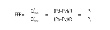
Fig. 1 The concept of fractional flow reserve (FFR). : hyperemic myocardial blood flow in the presence of a stenosis, : normal hyperemic myocardial blood flow, Pd: distal coronary pressure, Pa: aortic pressure, Pv: venous pressure, R: hyperemic myocardial resistance.

Fig. 2 Clinical application of FFR to a patient with multiple lesions and multi-vessel disease. By coronary angiography, 11 stenoses (arrow) were found, yet none of those in the left anterior descending and left circumflex arteries were functionally significant by FFR. FFR values measured in the right coronary artery was FFR 0.65 and pullback pressure tracing revealed the lesion at the mid right coronary artery (*) was hemodynamically the most significant stenosis. After one stent implantation at the mid right coronary artery, the FFR was 0.81. FFR: fractional flow reserve.
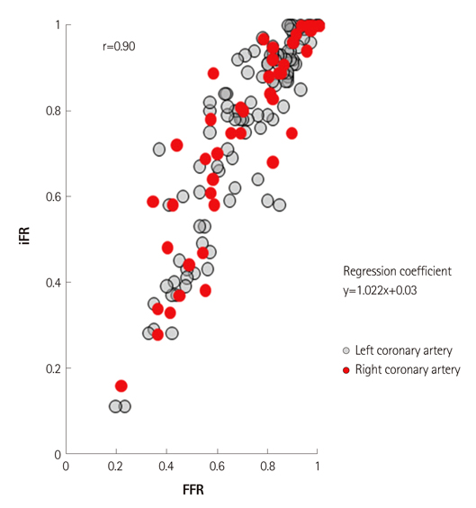
Fig. 3 Correlation between iFR and FFR according to the coronary artery (data from ADVISE study, courtesy of Justin Davies, MD). iFR: instantaneous wave-free ratio, FFR: fractional flow reserve.

Fig. 4 A case example of CT-derived computed FFR. By coronary CT angiography, significant stenosis was found at the proximal left anterior descending coronary artery. When this lesion was assessed by CT-derived computed FFR, FFRG was 0.74 and found to be functionally significant. This information derived from non-invasive assessment matched very well with invasive angiography and invasive FFR measurement (FFR=0.74). FFR: fractional flow reserve.

Fig. 5 Comparison of the diagnostic performance between FFRCT and CCTA (from DISCOVER FLOW study, per-vessel analysis, n=159). PPV: positive predictive value, NPV: negative predictive value, FFRCT: CT-derived computed FFR, CCTA: coronary CT angiography, DISCOVER FLOW: Diagnosis of Ischemia-Causing Stenoses Obtained Via Noninvasive Fractional Flow Reserve, FFR: fractional flow reserve.
Reference
-
1. Shaw LJ, Iskandrian AE. Prognostic value of gated myocardial perfusion SPECT. J Nucl Cardiol. 2004. 11:171–185.2. Shaw LJ, Berman DS, Maron DJ, et al. Optimal medical therapy with or without percutaneous coronary intervention to reduce ischemic burden: results from the Clinical Outcomes Utilizing Revascularization and Aggressive Drug Evaluation (COURAGE) Trial Nuclear Substudy. Circulation. 2008. 117:1283–1291.3. Tonino PA, De Bruyne B, Pijls NH, et al. Fractional flow reserve versus angiography for guiding percutaneous coronary intervention. N Engl J Med. 2009. 360:213–224.4. Pijls NH, van Schaardenburgh P, Manoharan G, et al. Percutaneous coronary intervention of functionally nonsignificant stenosis: 5-year follow-up of the DEFER Study. J Am Coll Cardiol. 2007. 49:2105–2111.5. White CW, Wright CB, Doty DB, et al. Does visual interpretation of the coronary arteriogram predict the physiologic importance of a coronary stenosis? N Engl J Med. 1984. 310:819–824.6. Vogel RA. Assessing stenosis significance by coronary arteriography: are the best variables good enough? J Am Coll Cardiol. 1988. 12:692–693.7. Koo BK, Park KW, Kang HJ, et al. Physiological evaluation of the provisional side-branch intervention strategy for bifurcation lesions using fractional flow reserve. Eur Heart J. 2008. 29:726–732.8. Tonino PA, Fearon WF, De Bruyne B, et al. Angiographic versus functional severity of coronary artery stenoses in the FAME Study fractional flow reserve versus angiography in multivessel evaluation. J Am Coll Cardiol. 2010. 55:2816–2821.9. Yong AS, Ng AC, Brieger D, Lowe HC, Ng MK, Kritharides L. Three-dimensional and two-dimensional quantitative coronary angiography, and their prediction of reduced fractional flow reserve. Eur Heart J. 2011. 32:345–353.10. Pijls NH, De Bruyne B, Peels K, et al. Measurement of fractional flow reserve to assess the functional severity of coronary-artery stenoses. N Engl J Med. 1996. 334:1703–1708.11. De Bruyne B, Bartunek J, Sys SU, Pijls NH, Heyndrickx GR, Wijns W. Simultaneous coronary pressure and flow velocity measurements in humans: feasibility, reproducibility, and hemodynamic dependence of coronary flow velocity reserve, hyperemic flow versus pressure slope index, and fractional flow reserve. Circulation. 1996. 94:1842–1849.12. Pijls NH, van Son JA, Kirkeeide RL, De Bruyne B, Gould KL. Experimental basis of determining maximum coronary, myocardial, and collateral blood flow by pressure measurements for assessing functional stenosis severity before and after percutaneous transluminal coronary angioplasty. Circulation. 1993. 87:1354–1367.13. De Bruyne B, Baudhuin T, Melin JA, et al. Coronary flow reserve calculated from pressure measurements in humans: validation with positron emission tomography. Circulation. 1994. 89:1013–1022.14. Bech GJ, Droste H, Pijls NH, et al. Value of fractional flow reserve in making decisions about bypass surgery for equivocal left main coronary artery disease. Heart. 2001. 86:547–552.15. Lindstaedt M, Yazar A, Germing A, et al. Clinical outcome in patients with intermediate or equivocal left main coronary artery disease after deferral of surgical revascularization on the basis of fractional flow reserve measurements. Am Heart J. 2006. 152:156.e1–156.e9.16. Potvin JM, Rodés-Cabau J, Bertrand OF, et al. Usefulness of fractional flow reserve measurements to defer revascularization in patients with stable or unstable angina pectoris, non-ST-elevation and ST-elevation acute myocardial infarction, or atypical chest pain. Am J Cardiol. 2006. 98:289–297.17. Fischer JJ, Wang XQ, Samady H, et al. Outcome of patients with acute coronary syndromes and moderate coronary lesions undergoing deferral of revascularization based on fractional flow reserve assessment. Catheter Cardiovasc Interv. 2006. 68:544–548.18. Berger A, Botman KJ, MacCarthy PA, et al. Long-term clinical outcome after fractional flow reserve-guided percutaneous coronary intervention in patients with multivessel disease. J Am Coll Cardiol. 2005. 46:438–442.19. Wongpraparut N, Yalamanchili V, Pasnoori V, et al. Thirty-month outcome after fractional flow reserve-guided versus conventional multivessel percutaneous coronary intervention. Am J Cardiol. 2005. 96:877–884.20. Smith SC Jr, Feldman TE, Hirshfeld JW Jr, et al. ACC/AHA/SCAI 2005 guideline update for percutaneous coronary intervention--summary article: a report of the American College of Cardiology/American Heart Association Task Force on Practice Guidelines (ACC/AHA/SCAI Writing Committee to Update the 2001 Guidelines for Percutaneous Coronary Intervention). Circulation. 2006. 113:156–175.21. Pijls NH, Fearon WF, Tonino PA, et al. Fractional flow reserve versus angiography for guiding percutaneous coronary intervention in patients with multivessel coronary artery disease: 2-year follow-up of the FAME (Fractional Flow Reserve Versus Angiography for Multivessel Evaluation) Study. J Am Coll Cardiol. 2010. 56:177–184.22. Fearon WF, Bornschein B, Tonino PA, et al. Economic evaluation of fractional flow reserve-guided percutaneous coronary intervention in patients with multivessel disease. Circulation. 2010. 122:2545–2550.23. Nam CW, Mangiacapra F, Entjes R, et al. Functional SYNTAX score for risk assessment in multivessel coronary artery disease. J Am Coll Cardiol. 2011. 58:1211–1218.24. Wijns W, Kolh P, Danchin N, et al. Guidelines on myocardial revascularization. Eur Heart J. 2010. 31:2501–2555.25. De Bruyne B, Pijls NH, Barbato E, et al. Intracoronary and intravenous adenosine 5'-triphosphate, adenosine, papaverine, and contrast medium to assess fractional flow reserve in humans. Circulation. 2003. 107:1877–1883.26. Wilson RF, Wyche K, Christensen BV, Zimmer S, Laxson DD. Effects of adenosine on human coronary arterial circulation. Circulation. 1990. 82:1595–1606.27. Kern MJ, Deligonul U, Tatineni S, Serota H, Aguirre F, Hilton TC. Intravenous adenosine: continuous infusion and low dose bolus administration for determination of coronary vasodilator reserve in patients with and without coronary artery disease. J Am Coll Cardiol. 1991. 18:718–729.28. Lindstaedt M, Bojara W, Holland-Letz T, et al. Adenosine-induced maximal coronary hyperemia for myocardial fractional flow reserve measurements: comparison of administration by femoral venous versus antecubital venous access. Clin Res Cardiol. 2009. 98:717–723.29. Seo MK, Shin DH, Yang HM, et al. Comparison of hyperemic efficacy between central and peripheral veous adenosine infusion for fractional flow reserve measurement. Circulation. 2010. 122:A18620. Abstract.30. Nair PK, Marroquin OC, Mulukutla SR, et al. Clinical utility of regadenoson for assessing fractional flow reserve. JACC Cardiovasc Interv. 2011. 4:1085–1092.31. Jang HJ, Koo BK, Kim JH, et al. Safety and efficacy of a novel hyperemic agent, nicorandil, for invasive physiologic assessment in a catheterization laboratory: a prospective multicenter study. Korean Circ J. 2011. 550608A. Abstract.32. Sen S, Escaned J, Malik IS, et al. Development and Validation of a New Adenosine-Independent Index of Stenosis Severity From Coronary Wave-Intensity Analysis: results of the ADVISE (ADenosine Vasodilator Independent Stenosis Evaluation) Study. J Am Coll Cardiol. 2012. 59:1392–1402.33. Kim HJ, Vignon-Clementel IE, Coogan JS, Figueroa CA, Jansen KE, Taylor CA. Patient-specific modeling of blood flow and pressure in human coronary arteries. Ann Biomed Eng. 2010. 38:3195–3209.34. Koo BK, Erglis A, Doh JH, et al. Diagnosis of ischemia-causing coronary stenoses by noninvasive fractional flow reserve computed from coronary computed tomographic angiograms. Results from the prospective multicenter DISCOVER-FLOW (Diagnosis of Ischemia-Causing Stenoses Obtained Via Noninasive Fractional Flow Reserve) study. J Am Coll Cardiol. 2011. 58:1989–1997.35. Min JK, Berman DS, Budoff MJ, et al. Rationale and design of the DeFACTO (Determination of Fractional Flow Reserve by Anatomic Computed Tomographic AngiOgraphy) Study. J Cardiovasc Comput Tomogr. 2011. 5:301–309.
- Full Text Links
-
- Actions
-
Cited
- CITED
-
- Close
- Share
-
- Similar articles
-
- Myocardial fractional flow reserve in acute myocardial infarction
- Funtional significance of the intermediate lesion in a single coronary artery assessed by fractional flow reserve
- Hemodynamic Significance of Coronary Cameral Fistula Assessed by Fractional Flow Reserve
- Future Directions in Coronary CT Angiography: CT-Fractional Flow Reserve, Plaque Vulnerability, and Quantitative Plaque Assessment
- A Case of Successful Percutaneous Coronary Intervention by Fractional Flow Reserve and 13N-Ammonia Positron Emission Tomography
