Four-Dimensional Real-Time Cine Images of Wrist Joint Kinematics Using Dual Source CT with Minimal Time Increment Scanning
- Affiliations
-
- 1Department of Radiology, Yonsei University College of Medicine, Seoul, Korea. hotsong@yuhs.ac
- KMID: 2158241
- DOI: http://doi.org/10.3349/ymj.2013.54.4.1026
Abstract
- PURPOSE
To validate the feasibility of real time kinematography with four-dimensional (4D) dynamic functional wrist joint imaging using dual source CT.
MATERIALS AND METHODS
Two healthy volunteers performed radioulnar deviation and pronation-supination wrist motions for 10 s and 4 s per cycle in a dual source CT scanner. Scan and reconstruction protocols were set to optimize temporal resolution. Cine images of the reconstructed carpal bone of the moving wrist were recorded. The quality of the images and radiation dosage were evaluated.
RESULTS
The 4D cine images obtained during 4 s and 10 s of radioulnar motion showed a smooth stream of movement with good quality and little noise or artifact. Images from the pronation-supination motion showed noise with a masked surface contour. The temporal resolution was optimized at 0.28 s.
CONCLUSION
Using dual source CT, 4D cine images of in vivo kinematics of wrist joint movement were obtained and found to have a shorter scan time, improved temporal resolution and lower radiation dosages compared with those previously reported.
MeSH Terms
Figure
-

Fig. 1 Optimized time increment for minimum temporal resolution. Time increment is defined as the time interval from the scan starting point of one series to that of the next series; in other words, the time gap between frames of four-dimensional motion images. Two examples of 1 s (A) and 0.30 s (B) time increments are shown here. The temporal resolution (0.28 s) is depicted as a square in the upper row and the time increment is depicted as an arrow in the lower row. The number of arrows is equal to the number of frames in the motion image. (A) With a 1 s time increment, fewer frames were obtained in the fixed scan time, and the data scanned during the remaining 0.72 s (0.28 s subtracted from 1 s) were not collected. (B) With a 0.30 s time increment, which is the closest value to the temporal resolution (0.28 s), more frames were obtained in the fixed scan time and the wasted data were only that scanned during 0.02 s (0.28 s subtracted from 0.30 s).
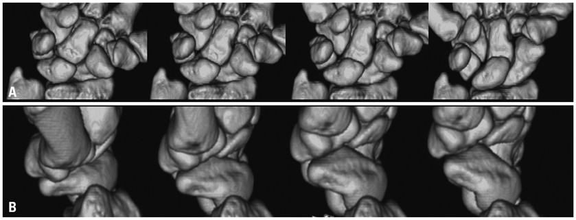
Fig. 2 Cine images of 10 s of radioulnar deviations per cycle from a 26-year-old healthy female volunteer: Coronal view (A) and sagittal view (B). Individual carpal bones are differentiated and their interactions are identified. Translocations of proximal carpal bones to the radial side and distal carpal bones to the ulnar side are shown in the coronal view. Multiplanar movement (extension) of the scaphoid along with radioulnar deviation is clearly depicted in the sagittal view.
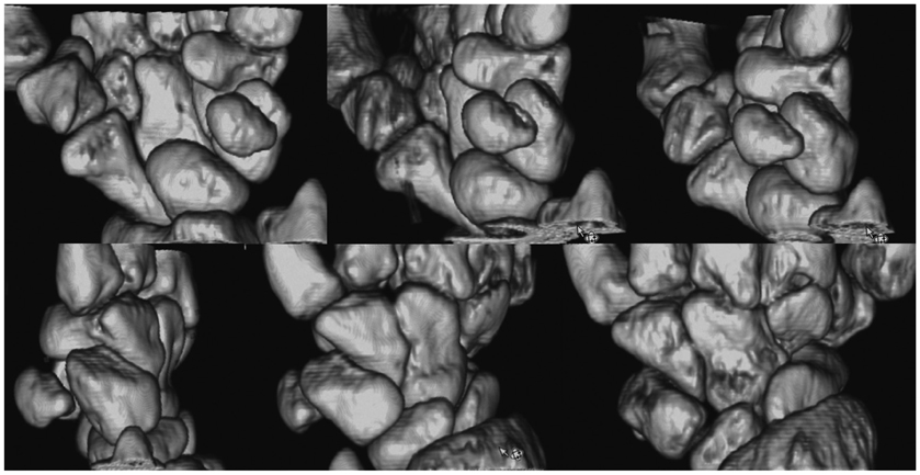
Fig. 3 Cine images of 10 s of wrist pronation-supination per cycle from a 27-year-old healthy female volunteer. Carpal bones and their interaction are depicted, but the image quality is lower than that of 10 s of radioulnar deviation per cycle due to motion artifact.
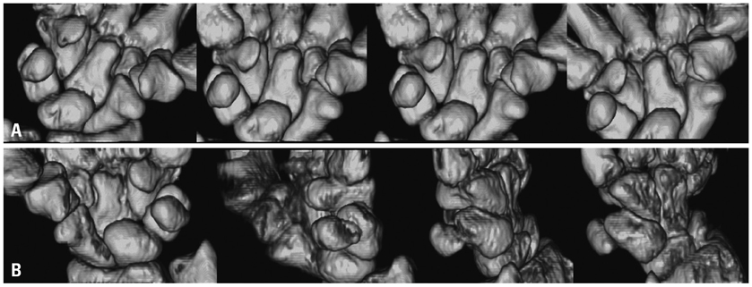
Fig. 4 Cine images of 4 s of radioulnar deviation (A) and pronation-supination (B) per cycle from a 27-year-old healthy female volunteer. With 4 s of radioulnar deviation per cycle, the margin and shape of the individual carpal bones are well visualized. The carpal bones are not fully covered in the 4 s radioulnar deviation images due to unintended forearm movement accompanying wrist movement, which is partially to reason that isolated wrist movement is more difficult during 4 s compared to 10 s. Though the pronation-supination images are lower quality than the radioulnar deviation images due to motion artifact, and the margin and shape of the carpal bones are blurred, the carpal bone movements are still depicted.
Reference
-
1. Toms AP, Chojnowski A, Cahir JG. Midcarpal instability: a radiological perspective. Skeletal Radiol. 2011; 40:533–541.
Article2. Carelsen B, Bakker NH, Strackee SD, Boon SN, Maas M, Sabczynski J, et al. 4D rotational x-ray imaging of wrist joint dynamic motion. Med Phys. 2005; 32:2771–2776.
Article3. Kobayashi M, Berger RA, Nagy L, Linscheid RL, Uchiyama S, Ritt M, et al. Normal kinematics of carpal bones: a three-dimensional analysis of carpal bone motion relative to the radius. J Biomech. 1997; 30:787–793.
Article4. Protas JM, Jackson WT. Evaluating carpal instabilities with fluoroscopy. AJR Am J Roentgenol. 1980; 135:137–140.
Article5. Jacobson JA, Oh E, Propeck T, Jebson PJ, Jamadar DA, Hayes CW. Sonography of the scapholunate ligament in four cadaveric wrists: correlation with MR arthrography and anatomy. AJR Am J Roentgenol. 2002; 179:523–527.
Article6. Finlay K, Lee R, Friedman L. Ultrasound of intrinsic wrist ligament and triangular fibrocartilage injuries. Skeletal Radiol. 2004; 33:85–90.
Article7. Chang W, Peduto AJ, Aguiar RO, Trudell DJ, Resnick DL. Arcuate ligament of the wrist: normal MR appearance and its relationship to palmar midcarpal instability: a cadaveric study. Skeletal Radiol. 2007; 36:641–645.
Article8. Mori S, Endo M, Obata T, Murase K, Fujiwara H, Susumu K, et al. Clinical potentials of the prototype 256-detector row CT-scanner. Acad Radiol. 2005; 12:148–154.9. Sun JS, Shih TT, Ko CM, Chang CH, Hang YS, Hou SM. In vivo kinematic study of normal wrist motion: an ultrafast computed tomographic study. Clin Biomech (Bristol, Avon). 2000; 15:212–216.
Article10. Tay SC, Primak AN, Fletcher JG, Schmidt B, Amrami KK, Berger RA, et al. Four-dimensional computed tomographic imaging in the wrist: proof of feasibility in a cadaveric model. Skeletal Radiol. 2007; 36:1163–1169.
Article11. Kalia V, Obray RW, Filice R, Fayad LM, Murphy K, Carrino JA. Functional joint imaging using 256-MDCT: technical feasibility. AJR Am J Roentgenol. 2009; 192:W295–W299.
Article12. Petersilka M, Bruder H, Krauss B, Stierstorfer K, Flohr TG. Technical principles of dual source CT. Eur J Radiol. 2008; 68:362–368.
Article13. Lichtman DM, Wroten ES. Understanding midcarpal instability. J Hand Surg Am. 2006; 31:491–498.
Article14. Berdia S, Short WH, Werner FW, Green JK, Panjabi M. The hysteresis effect in carpal kinematics. J Hand Surg Am. 2006; 31:594–600.
Article15. Wright TW, Dobyns JH, Linscheid RL, Macksoud W, Siegert J. Carpal instability non-dissociative. J Hand Surg Br. 1994; 19:763–773.
Article16. Schmitt R, Froehner S, Coblenz G, Christopoulos G. Carpal instability. Eur Radiol. 2006; 16:2161–2178.
Article17. Cerezal L, del Piñal F, Abascal F, García-Valtuille R, Pereda T, Canga A. Imaging findings in ulnar-sided wrist impaction syndromes. Radiographics. 2002; 22:105–121.
Article18. Watanabe A, Souza F, Vezeridis PS, Blazar P, Yoshioka H. Ulnar-sided wrist pain. II. Clinical imaging and treatment. Skeletal Radiol. 2010; 39:837–857.
Article19. Gardner MJ, Crisco JJ, Wolfe SW. Carpal kinematics. Hand Clin. 2006; 22:413–420.
Article20. Moore DC, Crisco JJ, Trafton TG, Leventhal EL. A digital database of wrist bone anatomy and carpal kinematics. J Biomech. 2007; 40:2537–2542.
Article21. Pfaeffle J, Blankenhorn B, Stabile K, Imbriglia J, Goitz R, Robertson D. Development and validation of a computed tomography-based methodology to measure carpal kinematics. J Biomech Eng. 2005; 127:541–548.
Article22. Crisco JJ, McGovern RD, Wolfe SW. Noninvasive technique for measuring in vivo three-dimensional carpal bone kinematics. J Orthop Res. 1999; 17:96–100.
Article23. Verdun FR, Bochud F, Gundinchet F, Aroua A, Schnyder P, Meuli R. Quality initiatives* radiation risk: what you should know to tell your patient. Radiographics. 2008; 28:1807–1816.
Article
- Full Text Links
-
- Actions
-
Cited
- CITED
-
- Close
- Share
-
- Similar articles
-
- Three-dimensional CT angiography of the canine hepatic vasculature
- Analysis of the Wrist Motion Using 3 - Dimensional Imaging
- Comparison of intraoral scanning and conventional impression techniques using 3-dimensional superimposition
- Retrospective Electrocardiography-Gated Real-Time Cardiac Cine MRI at 3T: Comparison with Conventional Segmented Cine MRI
- Usefulness of Two-dimensioanl CT & Three-dimensional CT in Blow-out Fracture
