Yonsei Med J.
2007 Aug;48(4):727-730. 10.3349/ymj.2007.48.4.727.
Granular Cell Tumors of the Abdominal Wall
- Affiliations
-
- 1Department of Pathology, Ansan Hospital, Medical College, Korea University, Ansan, Korea. repath@korea.ac.kr
- 2Department of General Surgery, Ansan Hospital, Medical College, Korea University, Ansan, Korea.
- KMID: 2158184
- DOI: http://doi.org/10.3349/ymj.2007.48.4.727
Abstract
- Granular cell tumors (GCT) are found in virtually any body site, including the tongue, skin, subcutaneous tissue, breast, rectum and vulva. However, they are rarely seen in the abdominal wall. We report here on a rare case of GCT in the rectus muscle of the abdominal wall. A 44-year-old woman presented with a non-tender, hard mass in the right lower abdominal wall. Upon microscopic examination, the tumor was found to comprise of large polygonal cells with an abundant eosinophilic granular cytoplasm and round to oval nuclei. Upon immunohistochemical staining, the large cells showed S-100 and CD68 positive granular aggregates in the cytoplasm. Many lysosomes of variable size were observed in the cytoplasm.
Keyword
MeSH Terms
Figure
-

Fig. 1 Upon gross examination, the cut surface of the mass shows a solid grayish-white appearance with focal fibrotic changes. This mass is poorly demarcated from the surrounding skeletal muscle tissue.
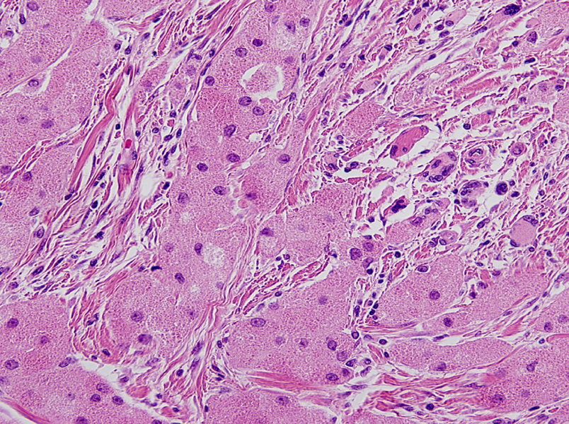
Fig. 2 Upon microscopic examination, the tumor is composed of large polygonal cells arranged in cord or sheets that are divided by slender fibrous tissue (H & E, × 400).
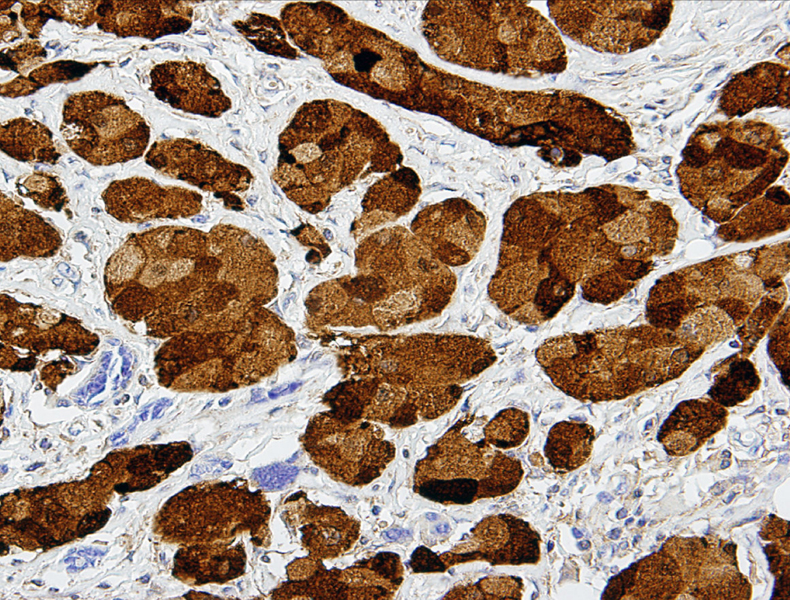
Fig. 3 Upon immunohistochemical staining, the granular cytoplasm is strongly reactive for S-100 protein (× 200).

Fig. 4 Ultrastructurally, the cytoplasm shows numerous membrane-bound vacuoles, presumably lysosomes, which are filled with electron-dense materials, such as fragmented rough endoplasmic reticulum and myelin components.
Reference
-
1. Abrikossoff A. Über myome, ausgehend von der quergestreiften willkrlichen muskulatur. Virchows Arch. 1926. 260:215–233.2. Fisher ER, Wechsler H. Granular cell myoblastoma-a misnomer. Electron microscopic and histochemical evidence concerning its Schwann cell derivation and nature (granular cell schwannoma). Cancer. 1962. 15:936–954.
Article3. Altavilla G, Brotto M, Busatto G, Boccu C, Ragni L. Granular cell tumor of the intrapancreatic common bile duct: one case report and review of the literature. Ultrastruct Pathol. 2004. 28:171–176.
Article4. Mazur MT, Shultz JJ, Myers JL. Granular cell tumor. Immunohistochemical analysis of 21 benign tumors and one malignant tumor. Arch Pathol Lab Med. 1990. 114:692–696.5. Fanburg-Smith JC, Meis-Kindblom JM, Fante R, Kindblom LG. Malignant granular cell tumor of soft tissue: diagnostic criteria and clinicopathologic correlation. Am J Surg Pathol. 1998. 22:779–794.6. Lack EE, Worsham GF, Callihan MD, Crawford BE, Klappenbach S, Rowden G, et al. Granular cell tumor: a clinicopathologic study of 110 patients. J Surg Oncol. 1980. 13:301–316.
Article7. Gorelkin L, Costantino MJ, Majmudar B. Granular cell tumor of the abdominal wall musculature. South Med J. 1978. 71:857–858.
Article8. Vamsy CM, Smile SR, Ratnakar CR, Veliath AJ. Malignant granular cell tumour. A case report and review of literature. Indian J Cancer. 1992. 29:31–33.9. Chelly I, Bellil K, Mekni A, Bellil S, Belhadjsalah M, Kchir N, et al. Malignant granular cell tumor of the abdominal wall. Pathologica. 2005. 97:130–132.10. Joshi AH, Aqel NM. Educational case report-self assessment. An anterior abdominal wall tumour. Cytopathology. 2003. 14:172221–222.11. Ordóñez NG, Mackay B. Granular cell tumor: a review of the pathology and histogenesis. Ultrastruct Pathol. 1999. 23:207–222.
Article12. Filie AC, Lage JM, Azumi N. Immunoreactivity of S100 protein, alpha-1-antitrypsin, and CD68 in adult and congenital granular cell tumors. Mod Pathol. 1996. 9:888–892.13. Lellé RJ, Park H, Brow CA. Benign granular cell tumor mimicking carcinoma of the breast. Report of a case. Eur J Gynaecol Oncol. 1992. 13:390–393.
- Full Text Links
-
- Actions
-
Cited
- CITED
-
- Close
- Share
-
- Similar articles
-
- Granular Cell Tumor Occurring at the Abdominal Wall: A Case Report
- Malignant Granular Cell Tumor of the Abdominal Wall Mimicking Desmoid Tumor: A Case Report with CT Imaging Findings and Literature Review
- Periurethral Granular Cell Tumor: A Case Report
- Granular Cell Tumor Occurring in the Chest Wall: A Case Report
- A Case of Granular Cell Tumors
