Yonsei Med J.
2005 Oct;46(5):710-714. 10.3349/ymj.2005.46.5.710.
A Case of Soft Tissue Myoepithelial Tumor Arising in Masticator Space
- Affiliations
-
- 1Department of Pathology, Dankook University College of Medicine, Chungnam, Korea. jaihyang@yahoo.co.kr
- KMID: 2158154
- DOI: http://doi.org/10.3349/ymj.2005.46.5.710
Abstract
- Soft tissue myoepithelial tumors of the head and neck region are very rare, and only one case of soft tissue myoepithelial tumor occurring in the masticator space has been reported in the world literature. A case of soft tissue myoepithelial tumor with benign histomorphology, but with an invasive growth pattern, occurred in the masticator space of a 46-year- old male patient. Magnetic resonance imaging of paranasal sinus/nasopharynx revealed a well-defined, lobulated heterogeneous mass with high signal intensity and dense calcification in the masticator space between the left mandible ramus and pterygoid process. Grossly, the tumor was a well- circumscribed ovoid solid mass and consisted of yellowish gray glistening firm tissue. Histologically, the tumor showed a multinodular growth pattern and consisted of epithelioid cells in chondromyxoid stroma and of spindle-shaped to ovoid cells in the hyaline stroma. The tumor cells appeared bland, and no mitosis or necrosis was found within the tumor. The tumor focally invaded to adhered bone tissue. Immunohistochemically, the tumor cells were diffusely positive for epithelial membrane antigen, smooth muscle actin, but negative for other epithelial markers. Ultrastructurally, the cytoplasm of the tumor cells contained sparse microfilaments and subplasmalemmal densities. Attenuated desmosomes were commonly seen between the tumor cells.
Keyword
MeSH Terms
Figure
-

Fig. 1 MR PNS nasopharynx revealed a well-defined, lobulated, heterogeneous mass with high signal intensity in the masticator space.
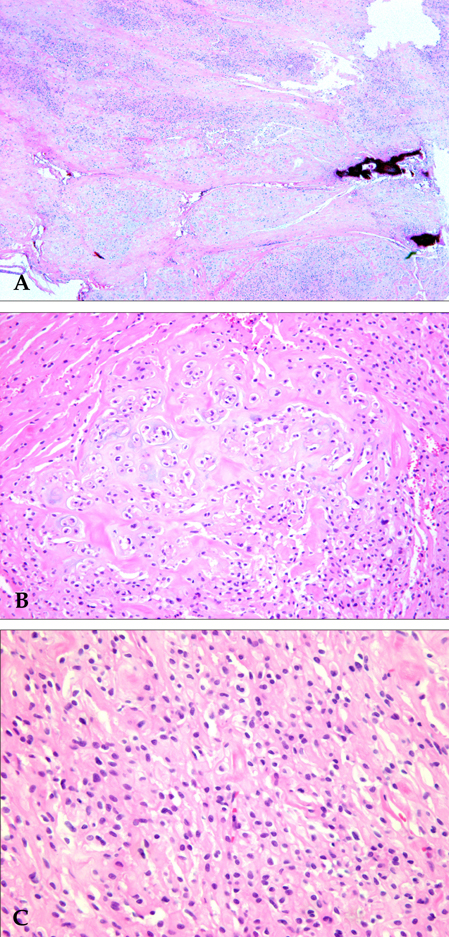
Fig. 2 Histologically, the tumor showed a multinodular growth pattern (A). The tumor was composed of epithelioid cells in the chondromyxoid stroma (B) and spindle-shaped to ovoid cells in the hyaline stroma (C). The tumor cells were cytologically bland-looking with no occurrence of mitosis.

Fig. 3 Immunohistochemically, the tumor cells were focally positive for EMA (A) and diffusely positive for smooth muscle actin (B).
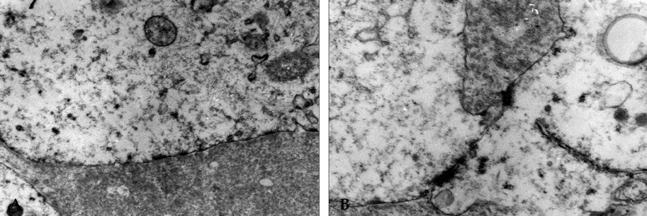
Fig. 4 Electron microscopic examination revealed cytoplasmic microfilaments and subplasmalemmal densities (×9000) (A) and attenuated desmosomes (×15000) (B).
Reference
-
1. Hornick JL, Fletcher CD. Myoepithelial tumors of soft tissue: a clinicopathologic and immunohistochemical study of 101 cases with evaluation of prognostic parameters. Am J Surg Pathol. 2003. 27:1183–1196.2. Kilpatrick SE, Hitchcock MG, Kraus MD, Calonje E, Fletcher CD. Mixed tumors and myoepitheliomas of soft tissue: a clinicopathologic study of 19 cases with a unifying concept. Am J Surg Pathol. 1997. 21:13–22.3. Minoda R, Masako M, Masuyama K, Yumoto E. Malignant myoepithelioma arising within the masticator space ectopically. Otolaryngol Head Neck Surg. 2001. 124:342–343.4. Kutzner H, Mentzel T, Kaddu S, Soares LM, Sangueza OP, Requena L. Cutaneous myoepithelioma: an under-recognized cutaneous neoplasm composed of myoepithelial cells. Am J Surg Pathol. 2001. 25:348–355.5. Mentzel T, Requena L, Kaddu S, Soares de Aleida LM, Sangueza OP, Kutzner H. Cutaneous myoepithelial neoplasms: clinicopathologic and immunohistochemical study of 20 cases suggesting a continuous spectrum ranging from benign mixed tumor of the skin to cutaneous myoepithelioma and myoepithelial carcinoma. J Cutan Pathol. 2003. 30:294–302.6. Hornick JL, Fletcher CD. Cutaneous myoepithelioma: a clinicopathologic and immunohistochemical study of 14 cases. Hum Pathol. 2004. 35:14–24.7. Bisceglia M, Cardone M, Fantasia L, Cenacchi G, Pasquinelli G. Mixed tumors, myoepitheliomas, and oncocytomas of the soft tissues are likely members of the same family: a clinicopathologic and ultrastructural study. Ultrastruct Pathol. 2001. 25:399–418.8. Smith BC, Ellis GL, Meis-Kindblom JM, Williams SB. Ectomesenchymal chondromyxoid tumor of the anterior tongue. Nineteen cases of a new clinicopathologic entity. Am J Surg Pathol. 1995. 19:519–530.9. Egerbacher M, Bock P. Lingual papillae. Am J Surg Pathol. 1997. 21:360.10. Sasaguri T, Tanimoto A, Arima N, Hamada T, Hashimoto H, Sasaguri Y. Myoepithelioma of soft tissue. Pathol Int. 1999. 49:571–576.
- Full Text Links
-
- Actions
-
Cited
- CITED
-
- Close
- Share
-
- Similar articles
-
- Masticator Space Lesions: MRI and CT Findings
- Dedifferentiated Extraskeletal Myxoid Chondrosarcoma of the Masticator Space: A Case Report
- Epithelial-myoepithelial Carcinoma Arising in the Nasal Cavity-Immunohistochemical and Electron Microscopic Study
- Myoepithelial Carcinoma Arising within an Adenomyoepithelioma of the Breast: A Case Report
- MR Imaging of Masticator Space Infection
