Myoepithelial Carcinoma Arising within an Adenomyoepithelioma of the Breast: A Case Report
- Affiliations
-
- 1Department of Radiology, Anam Hospital, Korea University College of Medicine, Seoul, Korea. krcho@korea.ac.kr
- 2Department of Radiology, Ansan Hospital, Korea University College of Medicine, Ansan, Korea.
- 3Department of Radiology, Guro Hospital, Korea University College of Medicine, Seoul, Korea.
- 4Department of Pathology, Anam Hospital, Korea University College of Medicine, Seoul, Korea.
- KMID: 2384732
- DOI: http://doi.org/10.3348/jksr.2017.77.1.27
Abstract
- Adenomyoepithelioma of the breast is a rare tumor. A myoepithelial carcinoma arising within an adenomyoepithelioma is even more unusual. There are a limited number of reports discussing myoepithelial carcinoma; most of them describe pathological findings, but not imaging findings. We present a case of a 55-year-old woman who had a screen-detected myoepithelial carcinoma arising within an adenomyoepithelioma in her right breast. Upon the completion of a mammography and sonography an oval shaped mass with an indistinct margin in the upper portion of the right breast had been seen. It as appeared to be a spiculated, irregular-shaped, peripheral-enhancing mass on an MRI. On sonography-guided biopsy, an epithelial-myothelial tumor was confirmed, and the possibility of myoepithelial carcinoma was suggested. Breast-conserving surgery with a sentinel lymph node dissection was performed, and a pathological examination revealed a myoepithelial carcinoma arising within an adenomyoepithelioma.
MeSH Terms
Figure
-
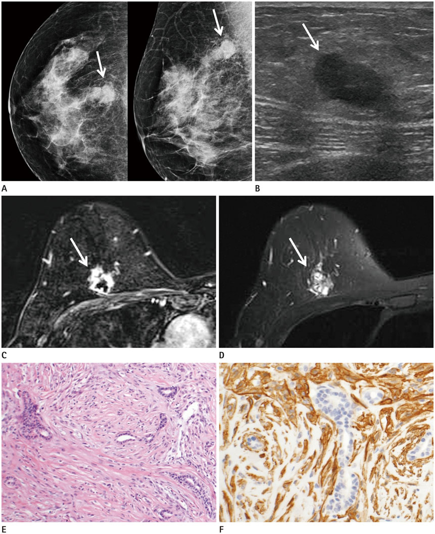
Fig. 1 A 55-year-old woman with myoepithelial carcinoma arising within adenomyoepithelioma. A. Screening mammography shows a 2 cm-sized, oval-shaped, isodense mass with indistinct margins (arrows) in the 12 o'clock direction of the right breast. B. Breast sonography shows an indistinct, oval, hypoechoic mass (arrow) seen in the right upper breast. There is no observed abnormal axillary lymphadenopathy. C. On MRI, axial fat-saturated T1-weighted subtraction image with post-contrast gadolinium injection shows an irregular mass with spiculated margins and rim enhancement (arrow) in the right upper mid-to-outer portion. D. On axial fat-saturated T2-weighted image, the mass was hyperintense (arrow). On dynamic study (not shown), this mass shows initial fast-enhancement and delayed plateau pattern. E. On histological examination (hematoxylin and eosin stain, × 100), the tumor shows a relatively uniform admixture of scattered, glandular, epithelial-lined spaces, and surrounding spindle and epithelioid myoepithelial-cell proliferation. F. On immunostaining (SMA, × 200), the myoepithelial cells are immunoreactive for SMA. SMA = smooth muscle actin
Reference
-
1. Ali RH, Hayes MM. Combined epithelial-myoepithelial lesions of the breast. Surg Pathol Clin. 2012; 5:661–699.2. American College of Radiology. ACR BI-RADS atlas: breast imaging reporting and data system. Reston, VA: American College of Radiology;2013.3. Howlett DC, Mason CH, Biswas S, Sangle PD, Rubin G, Allan SM. Adenomyoepithelioma of the breast: spectrum of disease with associated imaging and pathology. AJR Am J Roentgenol. 2003; 180:799–803.4. Endo Y, Sugiura H, Yamashita H, Takahashi S, Yoshimoto N, Iwasa M, et al. Myoepithelial carcinoma of the breast treated with surgery and chemotherapy. Case Rep Oncol Med. 2013; 2013:164761.5. Ruiz-Delgado ML, López-Ruiz JA, Eizaguirre B, Saiz A, Astigarraga E, Fernández-Temprano Z. Benign adenomyoepithelioma of the breast: imaging findings mimicking malignancy and histopathological features. Acta Radiol. 2007; 48:27–29.6. Petrozza V, Pasciuti G, Pacchiarotti A, Tomao F, Zoratto F, Rossi L, et al. Breast adenomyoepithelioma: a case report with malignant proliferation of epithelial and myoepithelial elements. World J Surg Oncol. 2013; 11:285.7. Adejolu M, Wu Y, Santiago L, Yang WT. Adenomyoepithelial tumors of the breast: imaging findings with histopathologic correlation. AJR Am J Roentgenol. 2011; 197:W184–W190.8. Tavassoli FA. Myoepithelial lesions of the breast. Myoepitheliosis, adenomyoepithelioma, and myoepithelial carcinoma. Am J Surg Pathol. 1991; 15:554–568.9. Moritz AW, Wiedenhoefer JF, Profit AP, Jagirdar J. Breast adenomyoepithelioma and adenomyoepithelioma with carcinoma (malignant adenomyoepithelioma) with associated breast malignancies: a case series emphasizing histologic, radiologic, and clinical correlation. Breast. 2016; 29:132–139.10. Park YM, Park JS, Jung HS, Yoon HK, Yang WT. Imaging features of benign adenomyoepithelioma of the breast. J Clin Ultrasound. 2013; 41:218–223.
