Yonsei Med J.
2005 Feb;46(1):61-65. 10.3349/ymj.2005.46.1.61.
The Diagnostic Value of Endoprobe for Small Esophageal Leiomyomas Derived from the Muscularis Mucosae
- Affiliations
-
- 1epartment of Internal Medicine, Institute of Gastroenterology, Yonsei University College of Medicine, Seoul, Korea. leeks519@yumc.yonsei.ac.kr
- KMID: 2158113
- DOI: http://doi.org/10.3349/ymj.2005.46.1.61
Abstract
- Esophageal leiomyoma derived from the muscularis mucosae (MM) is a rare condition, and the optimal modality for diagnosis and treatment is controversial. Endoscopic ultrasonography can provide an accurate image of esophageal layer structure, providing information on lesion suitability for potential endoscopic therapy. We attempted to investigate the diagnostic value of a transendoscopic balloon-tipped miniature ultrasonic endoprobe for small esophageal leiomyomas derived from MM. We resected 7 small esophageal leiomyomas derived from MM by endoscopic mucosal resection (EMR), all of which were diagnosed by a balloon-tipped endoprobe. The endosonographic and pathologic features of 7 cases of small esophageal leiomyomas derived from MM were compared. The balloon-tipped endoprobe clearly showed all 7 small esophageal leiomyomas derived from MM, even those under 5 mm in size (smallest lesion, 3.0 mm). The endosonographic characteristics of small esophageal leiomyomas derived from MM were a hypoechoic mass with smooth, regular, and a well-defined outer margin and homogenous inner echogram arising from the second hypoechoic layer. Complete resections were possible in all 7 cases by EMR without any complications. Tumor size was 3.0 - 13.5 mm (mean 7.8 mm) in maximum diameter. In all cases, endosonographic findings by endoprobe were exactly concordant with pathologic finding in determining the tumors depth in the esophageal wall, tissue origin and characteristics, growth pattern, and size. We detail the balloon-tipped endoprobe is a simple, convenient, and very useful in making accurate diagnosis of small esophageal leiomyomas derived from the MM and the appropriate applications of EMR.
Keyword
MeSH Terms
Figure
-
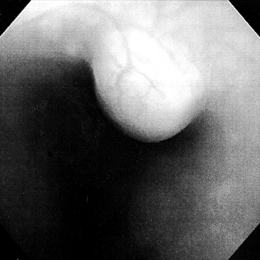
Fig. 1 Endoscopic finding of case No. 4 demonstrates a submucosal tumor with intact overlying mucosa.
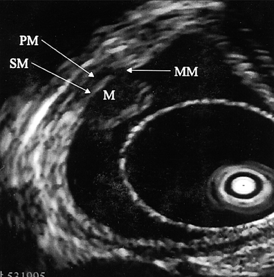
Fig. 2 High frequency ultrasonic endosonographic finding of case No. 4 shows a clear homogenous hypoechoic mass derived from the second layer of the esophagus (M, mass; MM, lamina muscularis mucosae; SM, submucosa; PM, proper muscle).

Fig. 3 Microscopic finding of resected specimens demonstrates the leiomyoma derived from the muscularis mucosae of the esophagus. Uniform, interlacing bundles of spindle-like cells without any mitotic bodies are seen (H&E stain, × 40).
Reference
-
1. Attah EB, Hajdu SI. Benign and malignant tumors of the esophagus at autopsy. J Thorac Cardiovasc Surg. 1968. 55:396–404.2. Massari M, Lattuada E, Zappa MA, Pieri G, Cioffi U, Simone MD, et al. Evaluation of leiomyoma of the esophagus with endoscopic ultrasonography. HepatoGastroenterology. 1997. 44:727–731.3. Kajiyama T, Sakai M, Torii A, Kishimoto H, Kin G, Uose S, et al. Endoscopic aspiration lumpectomy of esophageal leiomyomas derived from the muscularis mucosae. Am J Gastroenterol. 1995. 90(3):417. 422.4. Tio TL, Tytgat GNJ, den Hartog Jager FCA. Endoscopic ultrasonography for the evaluation of smooth muscle tumors in the upper gastrointestinal tract: an experience with 42 cases. Gastrointest Endosc. 1990. 36:342–350.5. Faivre J, Bory R, Moulinier B. Benign tumors of oesophagus: Value of endoscopy. Endoscopy. 1978. 10:264–268.6. Takada N, Higashino M, Osugi H, Tokuhara T, Kinoshita H. Utility of endoscopic ultrasonography in assessing the indications for endoscopic surgery of submucosal esophageal submucosal tumors. Surg Endosc. 1999. 13:228–230.7. Kojima T, Takahashi H, Parra-Blanco A, Kohsen K, Fujita R. Diagnosis of submucosal tumor of the upper GI tract by endoscopic resection. Gastrointest Endosc. 1999. 50:516–522.8. Solomon MP, Rosenblum H, Rosato FE. Leiomyoma of the esophagus. Ann Surg. 1984. 199:246–248.9. Jacobs WH, Bruns D. Endoscopic electrosurgical polypectomies of the upper gastrointestinal tract. Am J Gastroenterol. 1977. 68:241–248.10. Yu JP, Luo HS, Wang XZ. Endoscopic treatment of submucosal lesions of the gastrointestinal tract. Endoscopy. 1992. 24:190–193.11. Xu GQ, Zhang BL, Li YM, Chen LH, Ji F, Chen WX, et al. Diagnostic value of endoscopic ultrasonography for gastrointestinal leiomyoma. World J Gastroenterol. 2003. 9:2088–2091.12. Fleischer DE. Endoscopic resection of gastrointestinal tumors. Endoscopy. 1993. 25:479–481.
- Full Text Links
-
- Actions
-
Cited
- CITED
-
- Close
- Share
-
- Similar articles
-
- Intraluminal Pedunculated Leiomyoma in the Cervical Esophagus: Report of 1 Case
- Significance of Invasion to the Muscularis Mucosae on the Progression of Superficial Bladder Cancer
- Rectal Leiomyoma Diagnosed by Endoscopic Ultrasonography and Endoscopic Polypectomy
- An Esophageal Leiomyoma with Cystic Degeneration Mimicking a Malignant Neoplasm
- Clinical Outcomes of Endoscopic Removal in Patients with Colorectal Polypoid Leiomyomas
