An Esophageal Leiomyoma with Cystic Degeneration Mimicking a Malignant Neoplasm
- Affiliations
-
- 1Department of Internal Medicine, Pusan National University School of Medicine, Busan, Korea
- 2Biomedical Research Institute, Pusan National University Hospital, Busan, Korea
- 3Department of Pathology, Pusan National University Hospital, Busan, Korea
- KMID: 2547923
- DOI: http://doi.org/10.12771/emj.2023.e19
Abstract
- Esophageal subepithelial tumors (SETs) are commonly encountered during screening endoscopy, and leiomyomas are the most common SET of the esophagus. Almost all patients with esophageal leiomyomas are asymptomatic; however, some present with dysphagia, depending on the size of the tumor and the extent to which it encroaches on the lumen. The typical endosonographic features of esophageal leiomyomas include well-demarcated, homogeneously hypoechoic lesions with echogenicity similar to that of the surrounding proper muscle layer, but without cystic changes. Histopathologically, esophageal leiomyomas do not undergo cystic or myxoid degeneration. This report presents a case involving a 65-year-old man with a symptomatic esophageal SET and endosonographic features indicative of malignant neoplasms, who was diagnosed with esophageal leiomyoma with cystic and myxoid degeneration following surgical resection.
Figure
-
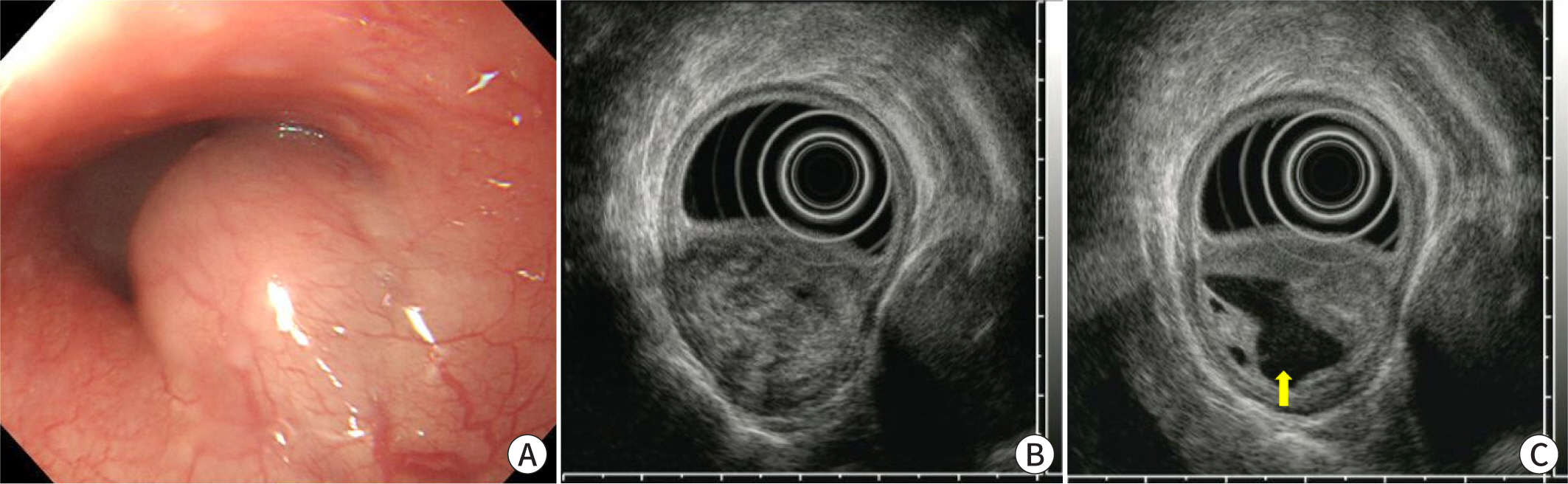
Fig. 1. Endoscopic and endosonographic findings. (A) A subepithelial tumor, measuring 3 cm, without surface erosion or ulceration is evident in the lower esophagus. (B, C) Endoscopic ultrasonography reveals a heterogeneously hypoechoic lesion with cystic changes (arrow) in the proper muscle layer.

Fig. 2. Contrast-enhanced chest CT. (A, B) Asymmetric wall thickening with heterogeneous enhancement and a non-enhanced area (arrow) is evident in the lower esophagus.
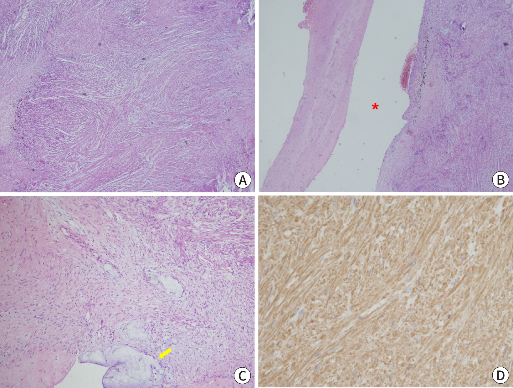
Fig. 3. Histopathological findings. (A) Intersecting bundles or fascicles of bland spindle cells with eosinophilic cytoplasm are observed (hematoxylin and eosin stain, ×40). (B, C) Cystic (asterisk) and myxoid degeneration (arrow) is evident; however, significant cytological atypia, increased mitotic activity, and tumor necrosis are not detected (hematoxylin and eosin stain, ×40 and ×100, respectively). (D) Tumor cells are positive for smooth muscle actin stain (×400).
Reference
-
References
1. Choe Y, Cho YK, Kim GH, Choi JH, Kim ES, Kim JH, et al. Prevalence, natural progression, and clinical practices of upper gastrointestinal subepithelial lesions in Korea: a multicenter study. Clin Endosc. 2023; Aug. 25. [Epub]. DOI: 10.5946/ce.2023.005. PMCID: PMC10244145.2. Lee MW, Kim GH. Diagnosing gastric mesenchymal tumors by digital endoscopic ultrasonography image analysis. Clin Endosc. 2021; 54((3)):324–328. DOI: 10.5946/ce.2020.061. PMID: 32549523. PMCID: PMC8182255.3. Kim DU, Kim GH, Ryu DY, Lee DG, Cheong JH, Lee BE, et al. Endosonographic features of esophageal granular cell tumors using a high-frequency catheter probe. Scand J Gastroenterol. 2011; 46((2)):142–147. DOI: 10.3109/00365521.2010.525661. PMID: 20950209.4. Seo WY, Kim GH. H I. An esophageal leiomyoma mistaken as an esophageal duplication cyst. Korean J Helicobacter Up Gastrointest Res. 2021; 21((2)):152–155. DOI: 10.7704/kjhugr.2020.0039.5. Kim GH, Park DY, Kim S, Kim DH, Kim DH, Choi CW, et al. Is it possible to differentiate gastric GISTs from gastric leiomyomas by EUS? World J Gastroenterol. 2009; 15((27)):3376–3381. DOI: 10.3748/wjg.15.3376. PMID: 19610138. PMCID: PMC2712898.6. Lee JS, Cho CM, Kwon YH, Seo AN, Bae HI, Han MH. Comparison of diagnostic performances of slow-pull suction and standard suction in endoscopic ultrasound-guided fine needle biopsy for gastrointestinal subepithelial tumors. Clin Endosc. 2022; 55((5)):637–644. DOI: 10.5946/ce.2021.257. PMID: 35973440. PMCID: PMC9539288.7. Shah P, Gao F, Edmundowicz SA, Azar RR, Early DS. Predicting malignant potential of gastrointestinal stromal tumors using endoscopic ultrasound. Dig Dis Sci. 2009; 54((6)):1265–1269. DOI: 10.1007/s10620-008-0484-7. PMID: 18758957.8. Ryu DG, Choi CW. Common gastric subepithelial tumors in Koreans. Korean J Helicobacter Up Gastrointest Res. 2022; 22((1)):29–37. DOI: 10.7704/kjhugr.2021.0051.9. Levine MS. Benign tumors of the esophagus. In. Gore RM, Levine MS, editors. editors. Textbook of gastrointestinal radiology. 2nd ed. Philadelphia: W.B. Saunders;2000. p. p. 387–402.10. Facciorusso A, Sunny SP, Del Prete V, Antonino M, Muscatiello N. Comparison between fine-needle biopsy and fine-needle aspiration for EUS-guided sampling of subepithelial lesions: a meta-analysis. Gastrointest Endosc. 2020; 91((1)):14–22. DOI: 10.1016/j.gie.2019.07.018. PMID: 31374187.11. Nakatani S, Okuwaki K, Watanabe M, Imaizumi H, Iwai T, Matsumoto T, et al. Stereomicroscopic on-site evaluation in endoscopic ultrasound-guided tissue acquisition of upper gastrointestinal subepithelial lesions. Clin Endosc. 2023; Apr. 18. [Epub]. DOI: 10.5946/ce.2022.288. PMID: 37070203.12. Fletcher CDM, Berman JJ, Corless C, Gorstein F, Lasota J, Longley BJ, et al. Diagnosis of gastrointestinal stromal tumors: a consensus approach. Hum Pathol. 2002; 33((5)):459–465. DOI: 10.1053/hupa.2002.123545. PMID: 12094370.13. Nabi Z, Reddy DN. Submucosal endoscopy: the present and future. Clin Endosc. 2023; 56((1)):23–37. DOI: 10.5946/ce.2022.139. PMID: 36617645. PMCID: PMC9902679.
- Full Text Links
-
- Actions
-
Cited
- CITED
-
- Close
- Share
-
- Similar articles
-
- A case of hemoperitoneum to spontaneous perforation of uterine leiomyoma with huge cystic degeneration
- An Esophageal Leiomyoma Mistaken as an Esophageal Duplication Cyst
- A Case of Huge Leiomyoma of the Broad Ligament with Secondary Cystic Degeneration
- A Case of Intraligamentary Leiomyoma with Huge Cystic Degeneration
- A Case of Esophageal Leiomyoma Showing High FDG Uptake on F-18 FDG PET
