Hypoplasia of Left Vertebral Artery with Intimal Fibromuscular Dysplasia in a Korean Woman
- Affiliations
-
- 1Department of Pathology, Jeju National University School of Medicine, Jeju, Korea.
- 2Department of Anatomy, Jeju National University School of Medicine, Jeju, Korea. spyoon@jejunu.ac.kr
- KMID: 2157929
- DOI: http://doi.org/10.3346/jkms.2012.27.7.811
Abstract
- We found a case of hypoplasia of vertebral artery with fibromuscular dysplasia in an 82-yr-old Korean female cadaver during a routine dissection course. In the present case, intracranial hypoplasia in left vertebral artery and bilateral origin of posterior inferior cerebellar artery at the vertebrobasilar junction were recognized. Histopathologically, left vertebral artery showed intimal type of fibromuscular dysplasia both in its extracranial and intracranial courses. These results indicate that the association of fibromuscular dysplasia and hypoplasia does exist in the vertebral artery, although the etiologies are not verified yet.
MeSH Terms
Figure
-
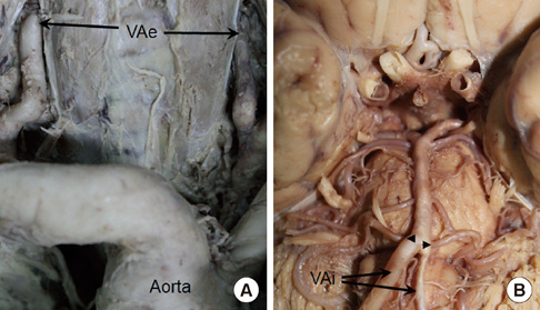
Fig. 1 Photographs of the vertebral arteries. Macroscopic hypoplasia was seen in left vertebral artery (VA), especially in the intracranial course. For histopathological classification, samples of the VA were obtained from its extracranial (VAe, A) and intracranial (VAi, B) course (arrows). Note the uncommon origin of the posterior inferior cerebellar arteries (arrowheads) from the basilar artery.

Fig. 2 Fibromuscular dysplasia of the vertebral artery (VA), intimal type. Right VA (A: hematoxylin and eosin, D: Orcein, G: anti-smooth muscle actin) was stained for control. Extracranial (VAe; B, E, H) and intracranial (VAi; C, F, I) left VA had stenotic lumen, marked fibrous thickening of tunica intima (TI), and disrupted internal elastic lamina (dotted arrows in E), while hyperplasia of smooth muscles in tunica media (TM) was not observed with anti-smooth muscle actin immunostaining (I). Neovascularization (asterisks) was also seen in left VAe with anti-CD31 immunostaining (rectangle from asterisk in B). Arrows, internal elastic lamina. Original magnification × 10 for A-C; × 40 for D-I.
Reference
-
1. Olin JW, Sealove BA. Diagnosis, management, and future developments of fibromuscular dysplasia. J Vasc Surg. 2011. 53:826–836.2. Touzé E, Oppenheim C, Trystram D, Nokam G, Pasquini M, Alamowitch S, Hervé D, Garnier P, Mousseaux E, Plouin PF. Fibromuscular dysplasia of cervical and intracranial arteries. Int J Stroke. 2010. 5:296–305.3. Songur A, Gonul Y, Ozen OA, Kucuker H, Uzun I, Bas O, Toktas M. Variations in the intracranial vertebrobasilar system. Surg Radiol Anat. 2008. 30:257–264.4. Min JH, Lee YS. Transcranial Doppler ultrasonographic evaluation of vertebral artery hypoplasia and aplasia. J Neurol Sci. 2007. 260:183–187.5. Macchi V, Porzionato A, Parenti A, De Caro R. The course of the posterior inferior cerebellar artery may be related to its level of origin. Surg Radiol Anat. 2004. 26:60–65.6. Kimura H, Hosoda K, Hara Y, Kohmura E. A very unusual case of fibromuscular dysplasia with multiple aneurysms of the vertebral artery and posterior inferior cerebellar artery. J Neurosurg. 2008. 109:1108–1112.7. Odero A, Bozzani A, Arici V, Aqozzino M. Hypoplasia and fibromuscular dysplasia of infrarenal abdominal aorta with downstream aneurysmmm: case report and review of the literature. J Vasc Surg. 2008. 48:1589–1592.8. Tan AK, Venketasubramanian N, Tan CB, Lee SH, Tjia TL. Ischaemic stroke from cerebral embolism in cephalic fibromuscular dysplasia. Ann Acad Med Singapore. 1995. 24:891–894.9. Kim DS, Kans SG, Yoo DS, Huh PW, Cho KS, Park CK. Ruptured right posterior inferior cerebellar artery aneurysm associated with hypoplasia of the right vertebral artery and both internal carotid arteries. Br J Neurosurg. 2009. 23:551–553.10. Schievink WI, Björnsson J. Fibromuscular dysplasia of the internal carotid artery: a clinicopathological study. Clin Neuropathol. 1996. 15:2–6.11. Barinagarrementeria F, Cantú Brito C, Izaguirre R, de la Peña A. Progressive intracranial occlusive disease associated with deficiency of protein S. Report of two cases. Stroke. 1993. 24:1752–1756.12. Chiche L, Bahnini A, Koskas F, Kieffer E. Occlusive fibromuscular disease of arteries supplying the brain: results of surgical treatment. Ann Vasc Surg. 1997. 11:496–504.
- Full Text Links
-
- Actions
-
Cited
- CITED
-
- Close
- Share
-
- Similar articles
-
- Pseudohypoplasia of Right Coronary Artery in a Korean Female Cadaver
- Fibromuscular Dysplasia of the Distal Internal Carotid and Middle Cerebral Artery
- A Case of Fibromuscular Dysplasia Associated with Cerebral Infarction
- Fibromuscular Dysplasia of the Middle Cerebal Artery
- Acute Renal Failure Due to Severe Stenosis of Fibromuscular Dysplasia in a Single Kidney
