Mesenchymal Hamartomas of the Liver: Comparison of Clinicopathologic Features between Cystic and Solid Forms
- Affiliations
-
- 1Department of Pathology, National Cancer Center, Seoul, Korea.
- 2Department of Pathology, Soonchunhyang University, Seoul, Korea.
- 3Department of Pathology, Yonsei University, Seoul, Korea. cppatholog@yumc.yonsei.ac.kr
- 4Department of Pathology, Seoul National University, Seoul, Korea.
- 5Department of Pathology, Sungkyungkwan University, Seoul, Korea.
- 6Department of Pathology, University of Ulsan, Seoul, Korea.
- 7Department of Pathology, Chungnam National University, Daegeon, Korea.
- 8Department of Pathology, Kyungpook National University, Daegu, Korea.
- KMID: 2157784
- DOI: http://doi.org/10.3346/jkms.2006.21.1.63
Abstract
- Mesenchymal hamartoma (MH) of the liver is an uncommon benign lesion related to ductal plate malformation. It is usually cystic and mainly composed of myxoid mesenchymal tissue with tortuous or cystic bile ducts. In order to characterize the clinicopathological features of MH, the Korean Gastrointestinal Pathology Study Group collected a total of 17 MH cases diagnosed in 7 hospitals from 1992 to 2002 and compared the clinicopathologic findings of cystic MH with those of solid variant. Among the 17 cases, 7 (41%) were solid. The solid form showed a higher serum level of alpha-fetoprotein (AFP), the smaller bile ducts, and more frequent proliferation of vessels. Serum AFP level was related to the amount of hepatocytes. Two of seven solid cases harbored a larger amount of evenly distributed hepatocytes and proliferation of small duct with focal hepatocyte-bile duct transition. These histologic findings are similar to those of mixed hamartoma. Therefore, the mixed hamartoma and the MH of both solid and cystic types could be the variants of one disease spectrum. And hepatocytes within MH might be rather a genuine tumor component than entrapped into the tumor. In conclusion, MH can show various clinicopathological features and recognition of these features will facilitate accurate diagnosis of MH.
Keyword
MeSH Terms
Figure
-
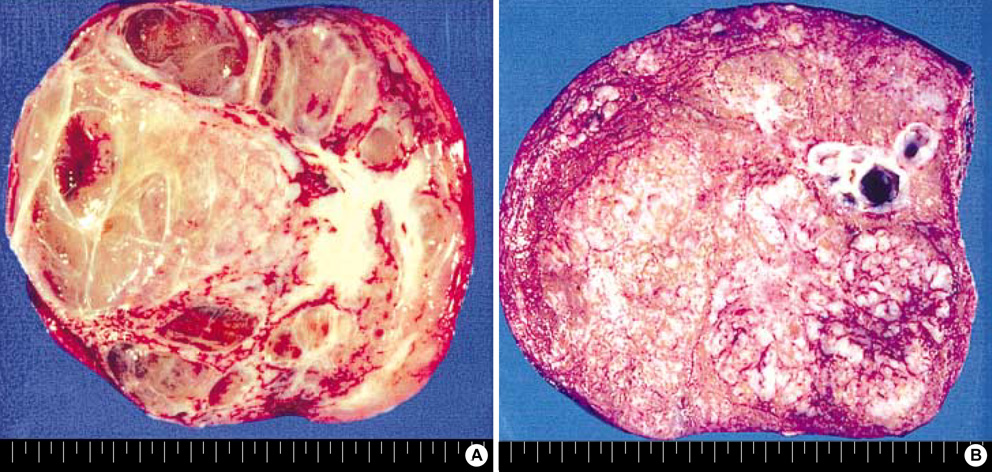
Fig. 1 Gross finding of MH. (A) Cystic MH (case 8). (B) Solid MH (case 7).

Fig. 2 Typical histologic feature of MH. (A) Histologic finding of adult case of MH, incidentally found in a 79-yr-old man (case 11) (H&E, ×40). (B) Myxoid mesenchymal stroma and proliferation of the architecturally abnormal bile ducts with periductal collaring of stromal cells (H&E, ×40).
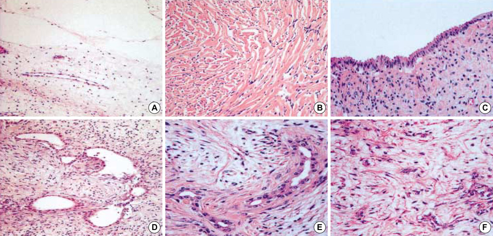
Fig. 3 Various histologic features of stroma and bile duct in MH. (A) Myxoid stroma (H&E, ×40). (B) Collagenous stroma (H&E, ×100). (C) Cystically dilated bile ducts (H&E, ×100). (D, E) Tortuous bile ducts (H&E, ×100 and ×200, respectively). (F) Small bile ducts (H&E, ×200).
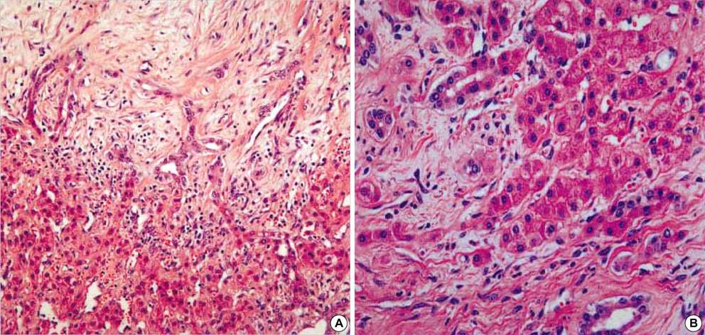
Fig. 4 Histologic finding of solid MH containing a larger amount of hepatocytes. (A) The bile ducts are ingrowing into hepatocytes (H&E, ×100). (B) Transition between hepatocytes and bile ducts is identified (H&E, ×200).
Reference
-
1. Stocker JT, Ishak KG. Mesenchymal hamartoma of the liver: report of 30 cases and review of the literature. Pediatr Pathol. 1983. 1:245–267.
Article2. Cook JR, Pfeifer JD, Dehner LP. Mesenchymal hamartoma of the liver in the adult: association with distinct clinical features and histological changes. Hum Pathol. 2002. 33:893–898.
Article3. Dehner LP, Ewing SL, Sumner HW. Infantile mesenchymal hamartoma of the liver. Histologic and ultrastructural observations. Arch Pathol. 1975. 99:379–382.4. Rakheja D, Margraf LR, Tomlinson GE, Schneider NR. Hepatic mesenchymal hamartoma with translocation involving chromosome band 19q13.4: a recurrent abnormality. Cancer Genet Cytogenet. 2004. 153:60–63.
Article5. Ramanujam TM, Ramesh JC, Goh DW, Wong KT, Ariffin WA, Kumar G, Taib NA. Malignant transformation of mesenchymal hamartoma of the liver: case report and review of the literature. J Pediatr Surg. 1999. 34:1684–1686.
Article6. Abdulkader I, Fraga M, Perez-Becerra E, Reyes-Santias RM, Bautista A, Chaves E, Forteza J. Mesenchymal hamartoma of the liver; Clinicopathological, immunohistochemical and flow cytometric study of two cases. Hepatol Res. 2004. 28:216–219.
Article7. DeMaioribus CA, Lally KP, Sim K, Isaacs H, Mahour GH. Mesenchymal hamartoma of the liver. A 35-year review. Arch Surg. 1990. 125:598–600.8. Sonobe H, Ohtsuki Y, Enzan H, Kurashige T, Matsuura K, Kaneko A, Ogata T. An unusual case of solid hamartoma in the liver. Acta Pathol Jpn. 1988. 38:75–82.
Article9. Ito H, Kishikawa T, Toda T, Arai M, Muro H. Hepatic mensenchymal hamartoma of an infant. J Pediatr Surg. 1984. 19:315–317.
Article10. Ohama K, Nagase H, Ogino K, Tsuchida K, Tanaka M, Kubo M, Horita S, Kawakami K, Ohmori M. Alpha-fetoprotein (AFP) levels in normal children. Eur J Pediatr Surg. 1997. 7:267–269.
Article11. Painter PC, Cope JY, Smith JI. Burtis CA, Ashwood ER, editors. Refence information for the clinical laboratory. Tietz textbook of clinical chemistry. 1999. Philadelphia: W.B. Saunders;1789–1840.12. Edmondson HA. Differential diagnosis of tumors and tumor-like lesions of liver in infancy and childhood. AMA J Dis Child. 1956. 91:168–186.
Article13. Rhodes RH, Marchildon MB, Luebke DC, Edmondson HA, Mikity VG. A mixed hamartoma of the liver: light and electron microscopy. Hum Pathol. 1978. 9:211–221.
Article14. Toda T, Yamada M, Nakama B, Yamazato M, Hokama A, Muto Y. Immunohistochemical and electron microscopic findings in a case of mixed hamartoma of the liver. Jpn J Surg. 1990. 20:101–106.
Article15. Chau KY, Ho JW, Wu PC, Yuen WK. Mesenchymal hamartoma of liver in a man: comparison with cases in infants. J Clin Pathol. 1994. 47:864–866.
Article16. Wada M, Ohashi E, Jin H, Nishikawa M, Shintani S, Yamashita M, Kano M, Yamanaka N, Nishigami T, Shimoyama T. Mesenchymal hamartoma of the liver: report of an adult case and review of the literature. Intern Med. 1992. 31:1370–1375.
Article17. Boman F, Bossard C, Fabre M, Diab N, Bonnevalle M, Boccon-Gibod L. Mesenchymal hamartomas of the liver may be associated with increased serum alpha foetoprotein concentrations and mimic hepatoblastomas. Eur J Pediatr Surg. 2004. 14:63–66.
Article
