Radiological Spectrum of Hepatic Mesenchymal Hamartoma in Children
- Affiliations
-
- 1Department of Radiology, Seoul National University College of Medicine and the Institute of Radiation Medicine, SNUMRC, Seoul, Korea. kimws@radcom.snu.ac.kr
- 2Department of Radiology, Sanggye Paik Hospital, Inje University, Seoul, Korea.
- 3Department of Radiology, Samsung Medical Center, Sungkyunkwan University School of Medicine, Seoul, Korea.
- 4Department of Pathology, Seoul National University College of Medicine, Seoul, Korea.
- KMID: 1089437
- DOI: http://doi.org/10.3348/kjr.2007.8.6.498
Abstract
- OBJECTIVE: A hepatic mesenchymal hamartoma is an uncommon benign tumor in children and little is known about the spectrum of its radiological features. The purpose of this study is to describe the spectrum of radiological features of a hepatic mesenchymal hamartoma in children. MATERIALS AND METHODS: Thirteen children with a pathologically confirmed hepatic mesenchymal hamartoma (M:F = 7:6; mean age, 3 years 2 months) were included in our study. Ultrasonography (US) was performed in nine patients including color and power Doppler US (n = 7). CT scans were performed in all patients. We evaluated the imaging findings of the hepatic mesenchymal hamartomas and the corresponding pathological features. RESULTS: Each patient had a single tumor (mean diameter: 13 cm [1.8-20 cm]). On CT and/or US, four patients (31%) had a "multiseptated cystic tumor", five patients (38%) had a " mixed solid and cystic tumor", and four patients (31%) had a "solid tumor." The septa of the cystic portion were thin in the multiseptated cystic tumors and irregularly thick in the mixed solid and cystic tumors as seen on US. On a post-contrast CT scan, solid portions or thick septa of the tumors showed heterogeneous enhancement. The amount of hepatocytes was significantly different among the three tumor groups according to the imaging spectrum (p = 0.042). CONCLUSION: A hepatic mesenchymal hamartoma in children can show a wide spectrum of radiological features, from a multiseptated cystic tumor to a mixed solid and cystic tumor, and even a solid tumor.
Keyword
MeSH Terms
-
Child
Child, Preschool
Contrast Media/administration & dosage
Female
Hamartoma/*diagnosis
Humans
Infant
Liver/*radiography/ultrasonography
Liver Neoplasms/*diagnosis
Male
Mesoderm/*radiography/ultrasonography
Observer Variation
Radiographic Image Enhancement/methods
Retrospective Studies
Tomography, X-Ray Computed/methods
Ultrasonography, Doppler, Color/methods
Figure
-
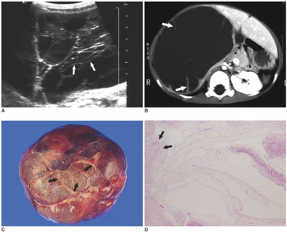
Fig. 1 A mesenchymal hamartoma in a 2-year-8-month-old boy (case 2). A. US shows a large, multiseptated cystic tumor in the right lobe of the liver. The septa of the tumor (arrows) are very thin and regular in thickness. B. A post-contrast CT scan shows a large cystic tumor with fine enhancing septa (arrows) in the liver. There is no solid portion or calcification within the tumor. C. A photograph of the gross specimen shows a huge tumor with a marked ballooning appearance and multiple septa (arrows). D. A photomicrograph shows variable sized cystic spaces with myxoid mesenchymal stroma and proliferation of the bile duct (arrows) along the septa. (Hematoxylin & Eosin staining, × 40)
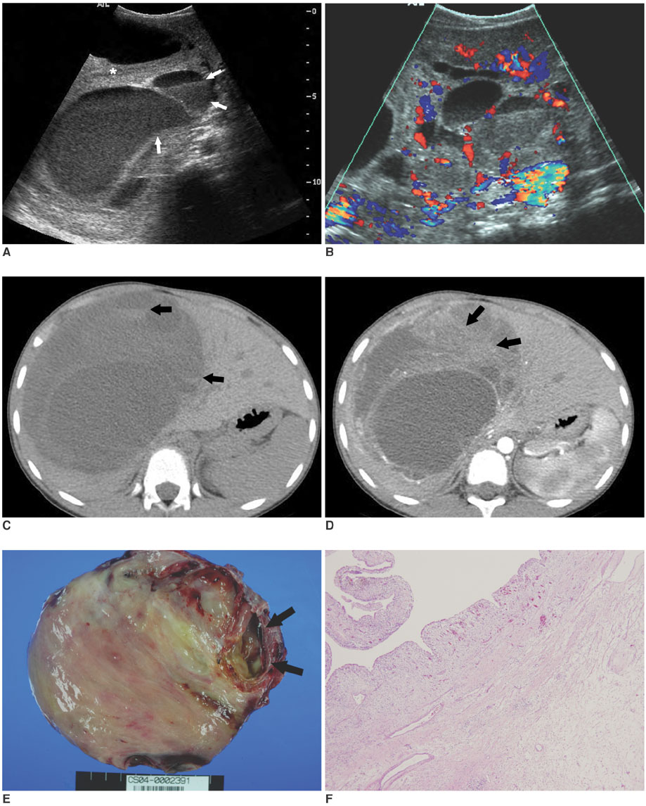
Fig. 2 A mesenchymal hamartoma in a 7-year-2-month-old boy (case 7). A. US shows a huge, mixed solid and cystic tumor. The echogenic materials and fluid-fluid levels (arrows) are noted in the cystic portions of the tumor. The solid portion of the tumor is hyperechoic (*). B. Color Doppler US shows vascularity along the thick septa and solid portion of the tumor. C. A pre-contrast CT scan shows a low attenuating tumor in the right lobe of the liver. Note the fluid-fluid levels due to an intracystic hemorrhage within some of the cystic areas (arrows). D. On a post-contrast CT scan, the solid portion of the tumor shows heterogeneous enhancement (arrows). E, F. A photograph of a cut section of the specimen shows a large tumor with a central solid portion and a peripheral cystic portion (arrows, E). The solid portion contains a large amount of myxoid mesenchymal stroma and proliferation of the bile duct, as seen on the photomicrograph (F, Hematoxylin & Eosin staining, × 40).

Fig. 3 A mesenchymal hamartoma in a 5-year-4-month-old girl (case 8). A. US shows a 3-cm-sized solid and cystic tumor (arrows) in the left lobe of the liver. B. A pre-contrast CT scan shows a tiny nodular calcification (arrow) at the peripheral portion of the tumor. C. A post-contrast CT scan shows a heterogeneously enhancing tumor (arrows) with focal cystic portions. D. A photograph of a cut section of the specimen shows variable sized cystic portions within the tumor.
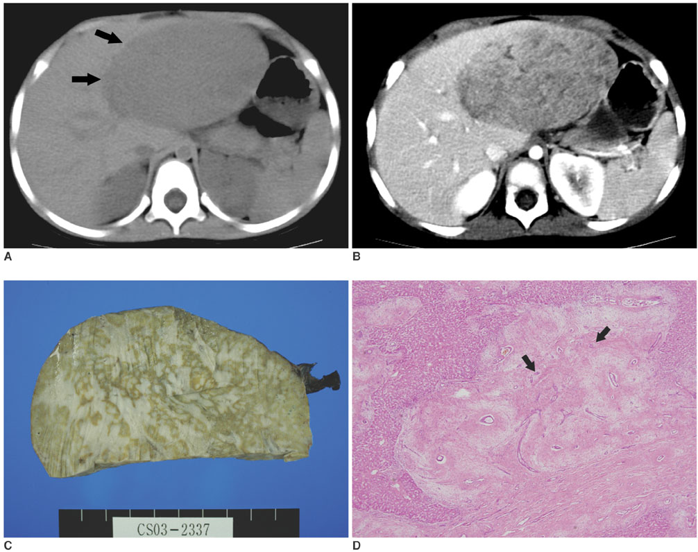
Fig. 4 A mesenchymal hamartoma in a 2-year-5-month-old girl (case 10). A. A pre-contrast CT scan shows a large, solid tumor (arrows) in the left lobe of the liver. The tumor shows low attenuation compared with the surrounding liver parenchyma on a pre-contrast CT. B. On a post-contrast CT scan, a heterogeneously enhancing tumor is seen in the left lobe of the liver. C. A photograph of the specimen shows a solid tumor with a marbling appearance. D. A photomicrograph shows large amount of myxoid mesenchymal stroma and proliferation of the bile duct (arrows). Hepatocytes are visible toward the left margin of the section (arrows, Hematoxylin & Eosin staining, × 40).
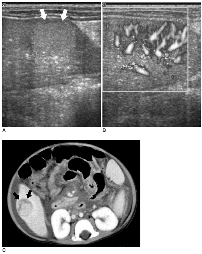
Fig. 5 A mesenchymal hamartoma in a 12-month-old girl that had a Kasai operation for biliary atresia (case 13). A. US shows a well-defined hyperechoic nodule (arrows) in the tip of the right lobe of the liver. B. Power Doppler US shows multiple intratumoral vascularities. C. On a post-contrast CT scan, a well-defined tumor is seen in the right lobe of the liver (arrows). The tumor shows a lobulated central portion with a peripheral rim of low attenuation. Histologically, the iso-attenuating portion contained a large amount of hepatocytes and a low attenuating rim of the peripheral portion revealed myxoid stroma. Ascites is seen.
Reference
-
1. Dehner LP, Ewing SL, Sumner HW. Infantile mesenchymal hamartoma of the liver. histologic and ultrastructural observations. Arch Pathol. 1975. 99:379–382.2. Stringer MD, Alizai NK. Mesenchymal hamartoma of the liver: a systematic review. J Pediatr Surg. 2005. 40:1681–1690.3. Ros PR, Goodman ZD, Ishak KG, Dachman AH, Olmsted WW, Hartman DS, et al. Mesenchymal hamartoma of the liver: radiologic-pathologic correlation. Radiology. 1986. 158:619–624.4. Federici S, Galli G, Sciutti R, Cuoghi D. Cystic mesenchymal hamartoma of the liver. Pediatr Radiol. 1992. 22:307–308.5. Stanley P, Hall TR, Woolley MM, Diament MJ, Gilsanz V, Miller JH. Mesenchymal hamartomas of the liver in childhood: sonographic and CT findings. AJR Am J Roentgenol. 1986. 147:1035–1039.6. Cetin M, Demirpolat G, Elmas N, Yüce G, Cetingül N, Balik E. Stromal predominant type mesenchymal hamartoma of liver: CT and MR features. Comput Med Imaging Graph. 2002. 26:167–169.7. Wada M, Ohashi E, Jin H, Nishikawa M, Shintani S, Yamashita M, et al. Mesenchymal hamartoma of the liver: report of an adult case and review of the literature. Intern Med. 1992. 31:1370–1375.8. Chung JH, Cho KJ, Choi DW, Lee BH, Chi JG. Solid mesenchymal hamartoma of the liver in adult. J Korean Med Sci. 1999. 14:335–337.9. Cook JR, Pfeifer JD, Dehner LP. Mesenchymal hamartoma of the liver in the adult: association with distinct clinical features and histological changes. Hum Pathol. 2002. 33:893–898.10. Edmondson HA. Differential diagnosis of tumors and tumor-like lesions of liver in infancy and childhood. AMA J Dis Child. 1956. 91:168–186.11. Helmberger TK, Ros PR, Mergo PJ, Tomczak R, Reiser MF. Pediatric liver neoplasms: a radiologic-pathologic correlation. Eur Radiol. 1999. 9:1339–1347.12. Stocker JT, Ishak KG. Mesenchymal hamartoma of the liver: report of 30 cases and review of the literature. Pediatr Pathol. 1983. 1:245–267.13. Alwaidh MH, Woodhall CR, Carty HT. Mesenchymal hamartoma of the liver: a case report. Pediatr Radiol. 1997. 27:247–249.14. Ito H, Kishikawa T, Toda T, Arai M, Muro H. Hepatic mesenchymal hamartoma of an infant. J Pediatr Surg. 1984. 19:315–317.15. Konez O, Goyal M, Vyas PK, Boinapally SB. Mesenchymal hamartoma of the liver. Comput Med Imaging Graph. 2001. 25:61–65.16. Chang HJ, Jin SY, Park C, Park YN, Jang JJ, Park CK, et al. Mesenchymal hamartoma of the liver: comparison of clinicopathologic features between cystic and solid forms. J Korean Med Sci. 2006. 21:63–68.17. Rhodes RH, Marchildon MB, Luebke DC, Edmondson HA, Mikity VG. A mixed hamartoma of the liver: light and electron microscopy. Hum Pathol. 1978. 9:211–221.18. Lack EE. Mesenchymal hamartoma of the liver. A clinical and pathologic study of nine cases. Am J Pediatr Hematol Oncol. 1986. 8:91–98.19. Okeda R. Mesenchymal hamartoma of the liver. An autopsy case with serial sections and some comments on its pathogenesis. Acta Pathol Jpn. 1976. 26:229–236.
