A Case of Sinusoidal Hemangioma with Lipoma
- Affiliations
-
- 1Department of Dermatology, Hallym University Sacred Heart Hospital, Anyang, Korea. dermakkh@yahoo.co.kr
- KMID: 2156802
- DOI: http://doi.org/10.5021/ad.2011.23.S2.S250
Abstract
- Sinusoidal hemangioma is a distinctive subset of a group of lesions known collectively as cavernous hemangiomas. Clinically, it develops in adults, predominantly females, and presents as a solitary, painless, bluish, deep dermal or subcutaneous nodule. Lipoma is the most common benign soft tissue tumor. Lipoma is distinguished from sinusoidal hemangioma on both clinical and histological grounds. Several studies have suggested that adipocytes originate from perivascular cells during adipogenesis. Angiogenic cytokines released by adipocytes play a role in the vasoproliferative response. The rearrangement or loss of chromosome 13 can also be associated with hemangioma. However, no previous cases of sinusoidal hemangioma have been associated with benign tumors like lipoma. Here, we describe an unusual case of sinusoidal hemangioma that occurred together with a lipoma on the right upper arm of a 43-year-old male.
MeSH Terms
Figure
-
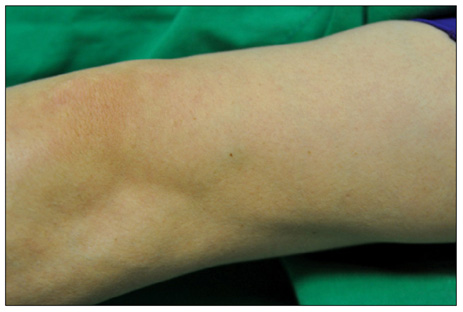
Fig. 1 A solitary, relatively well-defined, pea-sized, bluish subcutaneous nodule on the right upper arm.
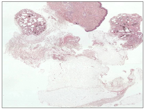
Fig. 2 On the upper left side of the picture, is a well-circumscribed, lobulated mass in the subcutaneous tissue. The mass is not encapsulated, and is composed of dilated and interconnecting vascular channels forming a sinusoidal structure. In the lower right region of the mass, is a well-circumscribed collection of benign mature adipocytes surrounded by a fibrous capsule (H&E, ×20).
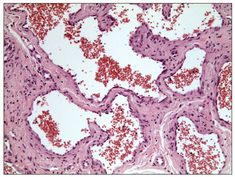
Fig. 3 Dilated vessels, with pseudopapillae and many red blood cells in the lumen, show a back-to-back arrangement (H&E, ×200).
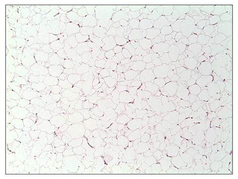
Fig. 4 In the lower right region of the hemangioma is, a benign tumor consisting of mature signet-ring fat cells (H&E, ×100).
Reference
-
1. Calonje E, Fletcher CD. Sinusoidal hemangioma. A distinctive benign vascular neoplasm within the group of cavernous hemangiomas. Am J Surg Pathol. 1991. 15:1130–1135.2. Nakamura M, Miyachi Y. Calcifying sinusoidal haemangioma on the back. Br J Dermatol. 1999. 141:377–378.
Article3. Kim EH, Kim YC, Lee SW. A case of polypoid sinusoidal hemangioma. Korean J Dermatol. 2006. 44:1016–1017.4. Ragsdale BD. Elder DE, Elenitsas R, Johnson BL, Murphy GF, Xu X, editors. Tumors with fatty, muscular, osseous, and/or cartilaginous differentiation. Lever's histopathology of the skin. 2009. 10th ed. Philadelphia: Lippincott Williams & Wilkins;1057–1106.5. Lee EJ, Oh EJ, Jeong TJ, Shin MK, Kim NI. A case of subcutaneous pyogenic granuloma, and a review of the literature. Korean J Dermatol. 2010. 48:776–779.6. Coras B, Hohenleutner U, Landthaler M, Hohenleutner S. Spindle cell hemangioma. Dermatol Surg. 2003. 29:875–878.
Article7. Kim SJ, Park HJ, Lee JY, Cho BK. A case of cellular angiolipoma. Korean J Dermatol. 2008. 46:1289–1291.8. Dotz W, Prioleau PG. Nevus lipomatosus cutaneus superficialis. A light and electron microscopic study. Arch Dermatol. 1984. 120:376–379.
Article9. Chang J, Most D, Bresnick S, Mehrara B, Steinbrech DS, Reinisch J, et al. Proliferative hemangiomas: analysis of cytokine gene expression and angiogenesis. Plast Reconstr Surg. 1999. 103:1–9.
Article10. Lucarelli E, Sangiorgi L, Benassi S, Donati D, Gobbi GA, Picci P, et al. Angiogenesis in lipoma: an experimental study in the chick embryo chorioallantoic membrane. Int J Mol Med. 1999. 4:593–596.
Article11. Dahlén A, Debiec-Rychter M, Pedeutour F, Domanski HA, Höglund M, Bauer HC, et al. Clustering of deletions on chromosome 13 in benign and low-malignant lipomatous tumors. Int J Cancer. 2003. 103:616–623.
Article12. Tharapel SA, Lewandowski RC, Tharapel AT, Wilroy RS Jr. Phenotype-karyotype correlation in patients trisomic for various segments of chromosome 13. J Med Genet. 1986. 23:310–315.
Article
