Ann Dermatol.
2011 Sep;23(Suppl 1):S139-S140. 10.5021/ad.2011.23.S1.S139.
Comment on "The Clinical Features and Pathophysiology of Acute Radiation Dermatitis in Patients Receiving Tomotherapy"
- Affiliations
-
- 1London Regional Lancer Program, London Health Sciences Centre, London, Ontario, Canada. slav.yartsev@lhsc.on.ca
- KMID: 2156773
- DOI: http://doi.org/10.5021/ad.2011.23.S1.S139
Abstract
- No abstract available.
MeSH Terms
Figure
-
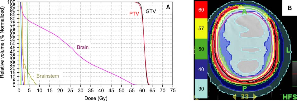
Fig. 1 (A) Cumulative relative dose-volume histogram and (B) dose distribution for the patient with large superficial target volume. GTV: gross tumor volume, PTV: planning target volume, unlabeled dose-volume histograms correspond to the right eye (purple), right lens (blue), right optic nerve (orange), optic chiasm (green), spinal cord (yellow).

Fig. 2 Clinical photographs of the patient's scalp: (A) before and (B) 2 months after tomotherapy treatment.
Reference
-
1. Lee JH, Kay CS, Maeng LS, Oh SJ, Lee AH, Lee JD, et al. The clinical features and pathophysiology of acute radiation dermatitis in patients receiving tomotherapy. Ann Dermatol. 2009. 21:358–363.
Article2. Hardcastle N, Soisson E, Metcalfe P, Rosenfeld AB, Tome WA. Dosimetric verification of helical tomotherapy for total scalp irradiation. Med Phys. 2008. 35:5061–5068.
Article3. Yukihara EG, McKeever SW. Optically stimulated luminescence (OSL) dosimetry in medicine. Phys Med Biol. 2008. 53:R351–R379.
Article
- Full Text Links
-
- Actions
-
Cited
- CITED
-
- Close
- Share
-
- Similar articles
-
- The Clinical Features and Pathophysiology of Acute Radiation Dermatitis in Patients Receiving Tomotherapy
- Helical Tomotherapy: Image-guided Intensity Modulated Radiation Therapy
- Analysis on the Calculated Dose in the Lung Radiation Surgery Planning Using TomoTherpay
- Clinical Significance of Helical Tomotherapy in Children with Extracranial Disease
- Helical Tomotherapy in Elderly Prostate Cancer Patients
