Korean J Neurotrauma.
2014 Apr;10(1):22-25. 10.13004/kjnt.2014.10.1.22.
Multiple Episodes of Hemorrhage Identified in MRI of Chronic Subdural Hematomas
- Affiliations
-
- 1Department of Neurosurgery, Soonchunhyang University Cheonan Hospital, Cheonan, Korea. ksleens@sch.ac.kr
- KMID: 2156093
- DOI: http://doi.org/10.13004/kjnt.2014.10.1.22
Abstract
OBJECTIVE
Septa within the hematoma cavity are common, especially in the mixed density chronic subdural hematomas (CSHs). Although CT remains the diagnosis of choice, MRI is superior to detect the membranes in CSHs. We could obtain MRIs in 64 patients with CSH. We examined the value of MRI to understand the history of CSH.
METHODS
We retrospectively examined the medical records and MRIs of 64 consecutive patients. MRI was selected to find any organic causes of neurologic symptoms. We classified the CSHs into septated or non-septated group, since classification of the septa was frequently obscure.
RESULTS
Septa were identified by MRI in 43 patients (67%). They were more common in the over 70-years-old group. Unknown causes were more common in the septated group, which implies they might suffer from multiple traumas. The signal intensity of the CSH was variable. The methods of treatment were different between two groups. Surgery was more common in the septated group (p=0.021). Surgery was performed in 57 patients (89%). Burr-hole drainage was successful in 55 patients, even in the septated group.
CONCLUSION
Septa within the hematoma cavity may be related to the multiple episodes of head trauma. Repeated trauma may cause acute bleedings over the CSHs, which is one of the pathogenic mechanisms of hematoma enlargement. MRI could show the history of CSH.
MeSH Terms
Figure
-
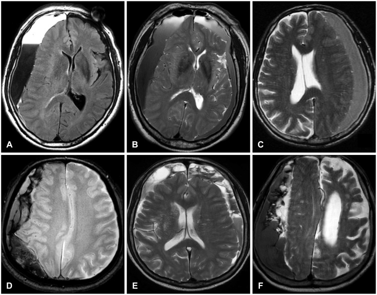
FIGURE 1 Examples of the non-septated and the septated groups. The layered (A) or graded (B) signal intensity is also classified into the non-septated group like the homogeneous (C) signal intensity. Since the patterns of the septa are usually mixed (D-F), it is hard to classify them into any specific subtypes.
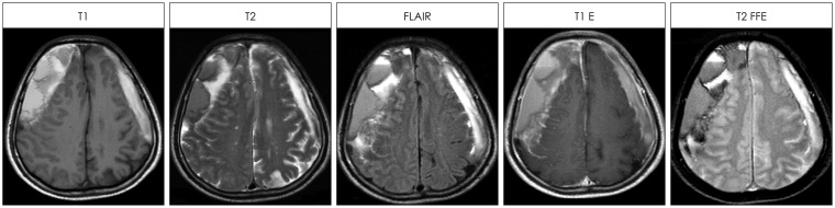
FIGURE 2 The signal intensity of the chronic subdural hematoma with septa was mixed one on T1-weighted, T2-weighted, fluid attenuated inversion recovery (FLAIR), T1-enhanced (T1 E) or T2 fast field echo (T2 FFE) images.
Cited by 1 articles
-
History of Chronic Subdural Hematoma
Kyeong-Seok Lee
Korean J Neurotrauma. 2015;11(2):27-34. doi: 10.13004/kjnt.2015.11.2.27.
Reference
-
1. Chon KH, Lee JM, Koh EJ, Choi HY. Independent predictors for recurrence of chronic subdural hematoma. Acta Neurochir (Wien). 2012; 154:1541–1548. PMID: 22653496.
Article2. Ducruet AF, Grobelny BT, Zacharia BE, Hickman ZL, DeRosa PL, Anderson K, et al. The surgical management of chronic subdural hematoma. Neurosurg Rev. 2012; 35:155–169. discussion 169. PMID: 21909694.
Article3. Fujisawa H, Nomura S, Kajiwara K, Kato S, Fujii M, Suzuki M. Various magnetic resonance imaging patterns of chronic subdural hematomas: indicators of the pathogenesis? Neurol Med Chir (Tokyo). 2006; 46:333–338. discussion 338-339. PMID: 16861826.
Article4. Gelabert-González M, Iglesias-Pais M, García-Allut A, Martínez-Rumbo R. Chronic subdural haematoma: surgical treatment and outcome in 1000 cases. Clin Neurol Neurosurg. 2005; 107:223–229. PMID: 15823679.
Article5. Hellwig D, Kuhn TJ, Bauer BL, List-Hellwig E. Endoscopic treatment of septated chronic subdural hematoma. Surg Neurol. 1996; 45:272–277. PMID: 8638225.
Article6. Hosoda K, Tamaki N, Masumura M, Matsumoto S, Maeda F. Magnetic resonance images of chronic subdural hematomas. J Neurosurg. 1987; 67:677–683. PMID: 3668635.
Article7. Imaizumi S, Onuma T, Kameyama M, Naganuma H. Organized ch-ronic subdural hematoma requiring craniotomy--five case reports. Neurol Med Chir (Tokyo). 2001; 41:19–24. PMID: 11218635.8. Kim JH, Kang DS, Kim JH, Kong MH, Song KY. Chronic subdural hematoma treated by small or large craniotomy with membranectomy as the initial treatment. J Korean Neurosurg Soc. 2011; 50:103–108. PMID: 22053228.
Article9. Krieg SM, Aldinger F, Stoffel M, Meyer B, Kreutzer J. Minimally invasive decompression of chronic subdural haematomas using hollow screws: efficacy and safety in a consecutive series of 320 cases. Acta Neurochir (Wien). 2012; 154:699–705. discussion 705. PMID: 22370998.
Article10. Le TH, Gean AD. Neuroimaging of traumatic brain injury. Mt Sinai J Med. 2009; 76:145–162. PMID: 19306377.
Article11. Lee JY, Ebel H, Ernestus RI, Klug N. Various surgical treatments of chronic subdural hematoma and outcome in 172 patients: is membranectomy necessary? Surg Neurol. 2004; 61:523–527. discussion 527-528. PMID: 15165784.
Article12. Lee KS, Shim JJ, Yoon SM, Doh JW, Yun IG, Bae HG. Acute-on-chronic subdural hematoma: not uncommon events. J Korean Neurosurg Soc. 2011; 50:512–516. PMID: 22323938.
Article13. Lo WL, Lee TC, Fang PS, Huang YH. Chronic subdural hematoma in patients under age 65 years: a comparative study of age cohort. Formosan J Surg. 2013; 46:10–14.
Article14. Markwalder TM. Chronic subdural hematomas: a review. J Neurosurg. 1981; 54:637–645. PMID: 7014792.
Article15. Nakaguchi H, Tanishima T, Yoshimasu N. Factors in the natural history of chronic subdural hematomas that influence their postoperative recurrence. J Neurosurg. 2001; 95:256–262. PMID: 11780895.
Article16. Park HR, Lee KS, Shim JJ, Yoon SM, Bae HG, Doh JW. Multiple densities of the chronic subdural hematoma in CT Scans. J Korean Neurosurg Soc. 2013; 54:38–41. PMID: 24044079.
Article17. Senturk S, Guzel A, Bilici A, Takmaz I, Guzel E, Aluclu MU, et al. CT and MR imaging of chronic subdural hematomas: a comparative study. Swiss Med Wkly. 2010; 140:335–340. PMID: 20349366.
- Full Text Links
-
- Actions
-
Cited
- CITED
-
- Close
- Share
-
- Similar articles
-
- Multiple Densities of the Chronic Subdural Hematoma in CT Scans
- Delayed Multiple Intracerebral Hemorrhage After Burr Hole Drainage of Chronic Subdural Hematoma During Failure of Warfarin Resumption in Atrial Fibrillation
- Magnetic Resonance Imaging in Severe Head Injury: Comparison with Computed Tomography
- Abusive Head Trauma in Infants and Children in Japan
- Infrequent Hemorrhagic Complications Following Surgical Drainage of Chronic Subdural Hematomas
