Clinical and Radiographic Outcomes of Simple Curettage and Graft Using Allogenic Bone or Bone Substitute for Enchondroma Involving Short Tubular Bone of the Hand and Foot
- Affiliations
-
- 1Department of Orthopaedic Sugary, Uijeongbu St. Mary's Hospital, School of Medicine, The Catholic University of Korea, Uijeongbu, Korea. wjbahk@cmc.cuk.ac.kr
- KMID: 2154353
- DOI: http://doi.org/10.4055/jkoa.2016.51.1.85
Abstract
- PURPOSE
We analyzed outcomes after management of enchondroma involving short tubular bones of the hand and foot by curettage and grafting using allogenic bone or bone substitutes.
MATERIALS AND METHODS
Twenty-two patients (allogenic bone 15 and bone substitutes 7 patients) were recruited. Clinical results were assessed by pain, cosmetic problem, range of motion of joint and the power of grasp. Radiographic outcomes were analyzed by degree of bone defect.
RESULTS
Clinically, 19 patients were classified as excellent and 3 patients as good. Three patients with K-wire fixation had pain with local irritation, which was easily controlled by removal of the K-wires. There were no complications including deep infection, delayed or nonunion, refracture. Radiographically, 20 cases were classified as group 1 (bone defect smaller than 3 mm) and the 2 remaining cases were classified as group 2 (bone defect 4-10 mm).
CONCLUSION
Curettage and graft using allogenic bone or bone substitute is an effective modality of treatment for enchondroma involving short tubular bones of the hand and foot. When combined with pathologic fracture, early surgical management could shorten duration of immobilization. Surgical management might be considered for the lesion involving the foot when discovered because of high incidence of pathologic fracture.
Keyword
MeSH Terms
Figure
-

Figure 1 Anatomic distribution of enchondromas in the hand and feet.
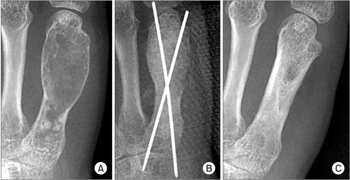
Figure 2 (A) Preoperative radiograph in a 37-year-old female shows a ballooning lytic lesion with intralesional calcification involving almost the entire 5th metacarpal bone. (B) Immediate postoperative radiograph demonstrates a lesion filled with allograft bone and fixed with K-wires. (C) A radiograph at 36 months after surgery shows a tiny defect less than 3 mm with normal cortex (group 1).

Figure 3 (A) Preoperative radiograph in a 21-year-old female shows a lytic lesion and intralesional calcification in association with a pathologic fracture involving 3/4 of the proximal phalanx of the middle finger. (B) Immediate postoperative radiograph shows a lesion filled with synthetic bone material (calcium phosphate) and fixed with K-wires. (C) A radiograph at 14 months after surgery shows no defect with normal cortex (group 1).
Reference
-
1. Fechner RE, Mills SE. Atlas of tumor pathology. Tumors of the bones and joints. Fascicle 8, 3rd series. Washington, DC: Armed Forces Institute of Pathology;1993. p. 86–112.2. Campanacci M. Bone and soft tissue tumors: clinical features, imaging, pathology and treatment. 2nd ed. Heidelberg: Springer;1999. p. 213–228.3. Casadei R, Ferraro A, Ferruzzi A, Biagini R, Ruggieri P. Bone tumors of the foot: epidemiology and diagnosis. Chir Organi Mov. 1991; 76:47–62.4. Grünert J, Strobel M, Brug E. Enchondroma of the hand. Z Orthop Ihre Grenzgeb. 1995; 133:180–186.5. Khan SN, Cammisa FP Jr, Sandhu HS, Diwan AD, Girardi FP, Lane JM. The biology of bone grafting. J Am Acad Orthop Surg. 2005; 13:77–86.
Article6. Finkemeier CG. Bone-grafting and bone-graft substitutes. J Bone Joint Surg Am. 2002; 84:454–464.
Article7. Younger EM, Chapman MW. Morbidity at bone graft donor sites. J Orthop Trauma. 1989; 3:192–195.
Article8. Bickels J, Wittig JC, Kollender Y, et al. Enchondromas of the hand: treatment with curettage and cemented internal fixation. J Hand Surg Am. 2002; 27:870–875.
Article9. Gaasbeek RD, Rijnberg WJ, van Loon CJ, Meyers H, Feith R. No local recurrence of enchondroma after curettage and plaster filling. Arch Orthop Trauma Surg. 2005; 125:42–45.
Article10. Joosten U, Joist A, Frebel T, Walter M, Langer M. The use of an in situ curing hydroxyapatite cement as an alternative to bone graft following removal of enchondroma of the hand. J Hand Surg Br. 2000; 25:288–291.
Article11. Kim JK, Kim NK. Curettage and calcium phosphate bone cement injection for the treatment of enchondroma of the finger. Hand Surg. 2012; 17:65–70.
Article12. Yasuda M, Masada K, Takeuchi E. Treatment of enchondroma of the hand with injectable calcium phosphate bone cement. J Hand Surg Am. 2006; 31:98–102.
Article13. Hung YW, Ko WS, Liu WH, et al. Local review of treatment of hand enchondroma (artificial bone substitute versus autologous bone graft) in a tertiary referral centre: 13 years' experience. Hong Kong Med J. 2015; 21:217–223.
Article14. Yercan H, Ozalp T, Coşkunol E, Ozdemir O. Long-term results of autograft and allograft applications in hand enchondromas. Acta Orthop Traumatol Turc. 2004; 38:337–342.15. Sassoon AA, Fitz-Gibbon PD, Harmsen WS, Moran SL. Enchondromas of the hand: factors affecting recurrence, healing, motion, and malignant transformation. J Hand Surg Am. 2012; 37:1229–1234.
Article16. Noble J, Lamb DW. Enchondromata of bones of the hand A review of 40 cases. Hand. 1974; 6:275–284.
Article17. Tordai P, Hoglund M, Lugnegrd H. Is the treatment of enchondroma in the hand by simple curettage a rewarding method? J Hand Surg Br. 1990; 15:331–334.
Article18. Goto T, Kawano H, Yamamoto A, et al. Simple curettage without bone grafting for enchondromas of the foot. Arch Orthop Trauma Surg. 2004; 124:301–305.
Article19. Sekiya I, Matsui N, Otsuka T, Kobayashi M, Tsuchiya D. The treatment of enchondromas in the hand by endoscopic curettage without bone grafting. J Hand Surg Br. 1997; 22:230–234.
Article20. Yanagawa T, Watanabe H, Shinozaki T, Takagishi K. Curettage of benign bone tumors without grafts gives sufficient bone strength. Acta Orthop. 2009; 80:9–13.
Article21. Hirn M, de Silva U, Sidharthan S, et al. Bone defects following curettage do not necessarily need augmentation. Acta Orthop. 2009; 80:4–8.
Article22. Hasselgren G, Forssblad P, Törnvall A. Bone grafting unnecessary in the treatment of enchondromas in the hand. J Hand Surg Am. 1991; 16:139–142.
Article23. Ablove RH, Moy OJ, Peimer CA, Wheeler DR. Early versus delayed treatment of enchondroma. Am J Orthop (Belle Mead NJ). 2000; 29:771–772.
- Full Text Links
-
- Actions
-
Cited
- CITED
-
- Close
- Share
-
- Similar articles
-
- Comparative Study of the Simple Curettage and the Curettage with Bonegraft in Enchondroma of the Hand
- Enchondroma of the Calcaneus: A Case Report
- A Case Report of Enchondroma on Right Ischjum
- Giant Cell Tumor of Short Tubular Bone: Case Report
- Early Single-Stage Curettage and Autogenous Bone Grafting for Enchondroma in the Hand with Minimally Displaced Pathologic Fracture
