MRI Findings of a Large Pedunculated Eccrine Poroma: A Case Report
- Affiliations
-
- 1Department of Radiology, Hanyang University Guri Hospital, Guri, Korea. ryuja@hanyang.ac.kr
- 2Department of Orthopedic Surgery, Hanyang University Guri Hospital, Guri, Korea.
- 3Department of Pathology, Hanyang University Guri Hospital, Guri, Korea.
- 4Department of Radiology, Hanyang University Hospital, Seoul, Korea.
- KMID: 2152606
- DOI: http://doi.org/10.3348/jksr.2016.74.2.132
Abstract
- Eccrine poroma is a rare benign neoplasm of the eccrine sweat gland that usually presents as a small skin lesion such as a papule or nodule. This benign tumor has an overall good prognosis; however, eccrine porocarcinomas can arise from long-standing pre-existing benign eccrine poromas. We reported the case of a 37-year-old man with mental retardation who presented with an 8-cm pedunculated and densely pigmented eccrine poroma on the left hip. The tumor showed low signal intensity on T1-weighted MRI, with inhomogenously high signal intensity on T2-weighted images and strong contrast enhancement after intravenous gadolinium administration. It directly extended from the dermal layer, and the subcutaneous tissue was preserved. Radiologists should be aware that eccrine poromas could be large and pedunculated. Furthermore, related MRI findings and diagnostic clues should be carefully considered.
MeSH Terms
Figure
-
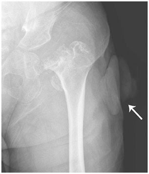
Fig. 1 Anterior-posterior radiograph shows a large pedunculated mass (arrow) in the left lateral hip. Severe osteolytic change of the left femoral head due to avascular necrosis is also seen.
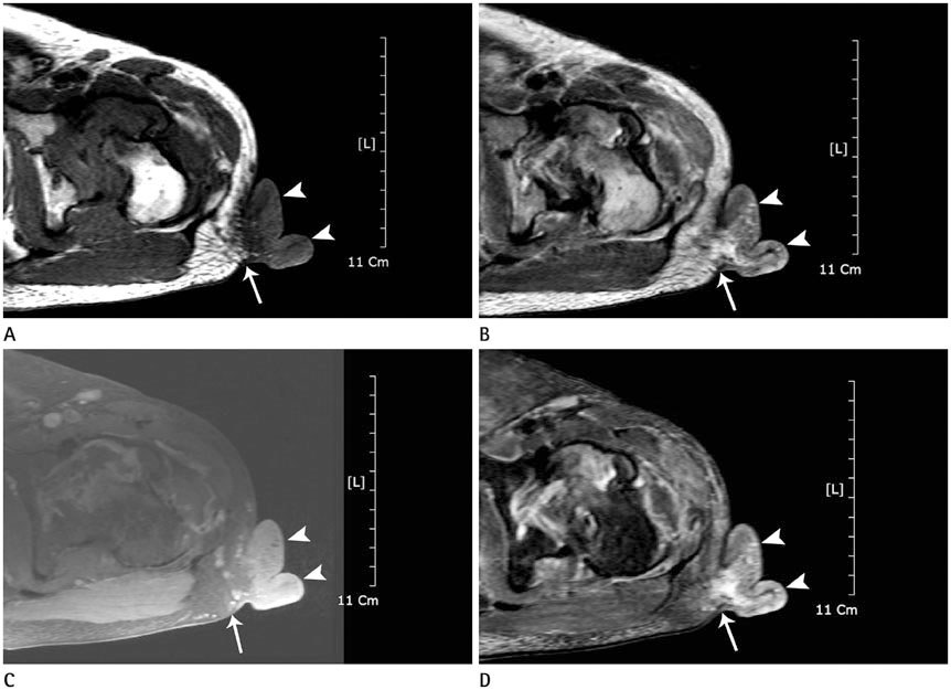
Fig. 2 Axial MRI of the pedunculated skin mass occupying dermal layer in the left lateral hip. A. On T1-weighted imaging, the mass extends from the dermal layer (arrow) and shows low signal intensity with subtle mottled low signal intensities in the periphery (arrowheads). B. On T2-weighted imaging, the mass extends from the dermal layer (arrow) shows heterogeneously high and low signal intensity with mottled high signal intensities in the periphery (arrowheads). C. On gadolinium-enhanced T1-weighted imaging, the mass shows strong enhancement with dilated, tortuous supplying and draining vessels in the base portion and underlying subcutaneous fat layer (arrow). The peripheral mottled lesions show low signal intensity without enhancement (arrowheads). D. On fat-suppressed T2-weighted imaging, the mass shows heterogeneously high signal intensity. Small portion of tumor infiltration is noted at the subcutaneous fat layer (arrow). Mottled high signal intensities are observed in the periphery (arrowheads).

Fig. 3 Gross and microscopic findings with histopathologic features. A. A dome-shaped exophytic mass with a tan brown color and surface nodularity. B. The cut section reveals a multilobulated, tan-white, soft mass occupying the dermis layer. C, D. Histologically, the lesion is composed of a proliferation of uniform basaloid cells with broad columns and cord arrangement with ductal structures lined by cuticles interspersed [hematoxylin-eosin stain, × 12.5 (C), × 200 (D)].
Reference
-
1. Goldman P, Pinkus H, Rogin JR. Eccrine poroma; tumors exhibiting features of the epidermal sweat duct unit. AMA Arch Derm. 1956; 74:511–521.2. Kang MC, Kim SA, Lee KS, Cho JW. A case of an unusual eccrine poroma on the left forearm area. Ann Dermatol. 2011; 23:250–253.3. Vu PP, Whitehead KJ, Sullivan TJ. Eccrine poroma of the eyelid. Clin Experiment Ophthalmol. 2001; 29:253–255.4. Iannicelli E, Galluzzo A, Salvi PF, Ziparo V, David V. A large porocarcinoma of perineal region: MR findings and review of the literature. Abdom Imaging. 2008; 33:744–747.5. Cunningham NG, Crockett RS, Kushner D. Intraepidermal eccrine adenocarcinoma of the foot: a case report. Foot Ankle Online J. 2009; [Epub]. DOI: 10.3827/faoj.2009.0207.0001.6. Leijs MM, Merk HF, Megahed M. [Eccrine poroma]. Hautarzt. 2013; 64:328–329.7. Horie K, Ito K, Hirata Y, Ito M. Eccrine poroma on the helix: a rare anatomical presentation. Clin Exp Dermatol. 2015; 40:442–444.8. Allende I, Gardeazabal J, Acebo E, Díaz-Pérez JL. [Pigmented eccrine poroma]. Actas Dermosifiliogr. 2008; 99:496–498.9. Cárdenas ML, Díaz CJ, Rueda R. Pigmented eccrine poroma in abdominal region, a rare presentation. Colomb Med (Cali). 2013; 44:115–111.
- Full Text Links
-
- Actions
-
Cited
- CITED
-
- Close
- Share
-
- Similar articles
-
- A Case of Pedunculated Pigmented Eccrine Poroma Combined with Congenital Melanocytic Nevus on the Scalp
- A Case of Eccrine Poroepithelioma
- Eccrine Poroma with Rapid Growth during Pregnancy: A Case Report and Review of the Literature
- A Case of Malignant Eccrine Poroma
- A Case of Eccrine Poroma with A Large Cystic Space
