Metastatic Brain Neuroendocrine Tumor Originating from the Liver
- Affiliations
-
- 1Department of Internal Medicine, The Catholic University of Korea, Bucheon St. Mary's Hospital, Bucheon, Korea.
- 2Department of Neurosurgery, The Catholic University of Korea, Bucheon St. Mary's Hospital, Bucheon, Korea. jkw94@naver.com
- 3Department of Hospital Pathology, The Catholic University of Korea, Bucheon St. Mary's Hospital, Bucheon, Korea.
- 4Department of Surgery, The Catholic University of Korea, Bucheon St. Mary's Hospital, Bucheon, Korea.
- KMID: 2151158
- DOI: http://doi.org/10.3340/jkns.2015.58.6.550
Abstract
- A 67-year-old male presented with left temporal hemianopsia and left hemiparesis. A contrast-enhanced magnetic resonance image revealed a 4.5x3.5x5.0 cm rim-enhancing mass with central necrosis and associated edema located in the left occipital lobe. Of positron emission tomography and abdominal computed tomography, a 9-cm mass with poor enhancement was found in the right hepatic lobe. Craniotomy and right hemihepatectomy was performed. The resected specimen showed histological features and immunochemical staining consistent with a metastatic neuroendocrine tumor (NET). Four months later, the tumors recurred in the brain, liverand spinal cord. Palliative chemotherapy with etoposide and cisplatin led to complete remission of recurred lesions, but the patient died for pneumonia. This is the first case of a metastatic brain NET originating from the liver. If the metastatic NET of brain is suspicious, investigation for primary lesion should be considered including liver.
Keyword
MeSH Terms
Figure
-
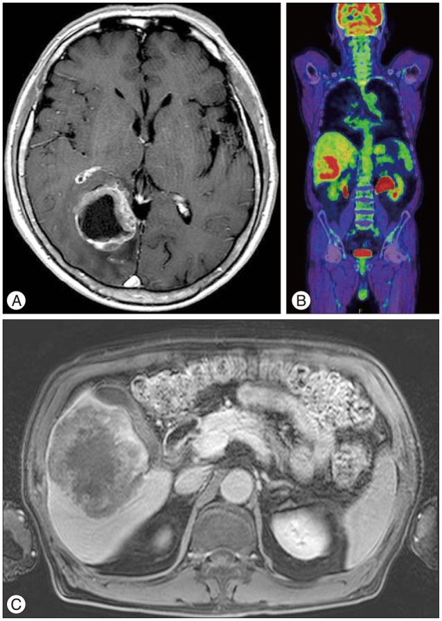
Fig. 1 A : Axial, T1-weighted, post-gadolinium image of the brain. B : Positron emission tomography-computed tomography (PET-CT). C : Axial, T1-weighted, post-gadolinium image of the liver. Note a rim-enhancing mass with central necrosis and associated edema in the right occipital lobe of brain and right hepatic lobe.
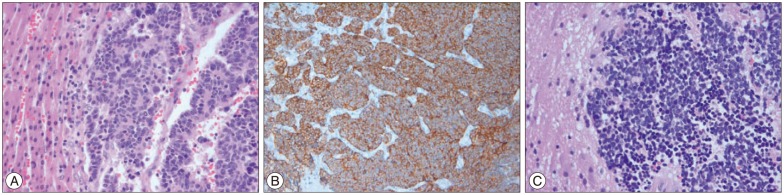
Fig. 2 Histopathologic findings of brain and liver mass show a round to oval stippled and scant, pink dranular cytoplasm with H&E (A and B) and brain mass are strongly stained with CD56 (C). This finding reveals that brain mass is a neuroendocrine tumor which is the same pathology with liver tissue.
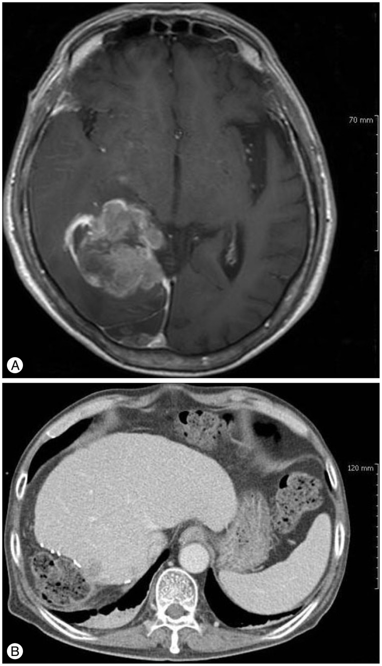
Fig. 3 A : Axial, T1-weighted, post-gadolinium image of the brain. B : Abdomen enhanced CT at 4 months after first craniotomy and hepatectomy. Note suspicious recurrence in the occipital lobe, inferior right cerebellum, and segment 8 of the liver.

Fig. 4 A : PET-CT scan after whole-brain radiation therapy. Note the multifocal FDG uptake in the cervical, thoracic and lumbar spinal cord and malignant tumor in right hepatectomy state. B : Spine post-gadolinium image. Note metastasis along the meninges at the T12–L1 level and cauda equine along the lumbar spine.
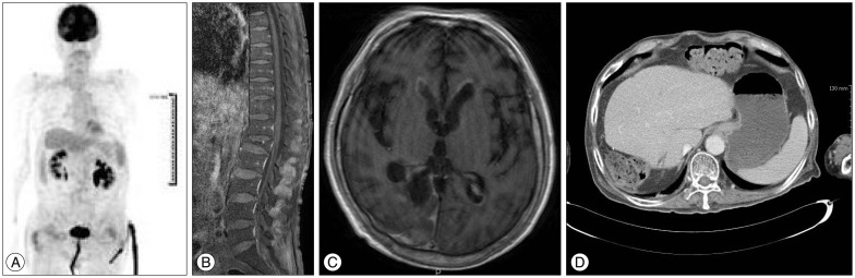
Fig. 5 A : PET-CT. B : spine post-gadolinium image. C : T1-weighted, post-gadolinium image of the brain. D : Abdomen enhanced CT. Note that previously enhancing tumors have disappeared.
Cited by 1 articles
-
젊은 나이에 발생하는 원발성 간 신경내분비 종양 1예
Sanghwa Song, Yangseok Koh
Korean J Gastroenterol. 2022;79(1):35-40. doi: 10.4166/kjg.2021.139.
Reference
-
1. Akahoshi T, Higashi H, Tsuruta S, Tahara K, Matsumoto T, Takeuchi H, et al. Primary neuroendocrine carcinoma coexisting with hemangioma in the liver : report of a case. Surg Today. 2010; 40:185–189. PMID: 20107963.
Article2. Burger PC, Green SB. Patient age, histologic features, and length of survival in patients with glioblastoma multiforme. Cancer. 1987; 59:1617–1625. PMID: 3030531.
Article3. Donadon M, Torzilli G, Palmisano A, Del Fabbro D, Panizzo V, Maggioni M, et al. Liver resection for primary hepatic neuroendocrine tumours : report of three cases and review of the literature. Eur J Surg Oncol. 2006; 32:325–328. PMID: 16426802.
Article4. Hlatky R, Suki D, Sawaya R. Carcinoid metastasis to the brain. Cancer. 2004; 101:2605–2613. PMID: 15495181.
Article5. Huang YQ, Xu F, Yang JM, Huang B. Primary hepatic neuroendocrine carcinoma : clinical analysis of 11 cases. Hepatobiliary Pancreat Dis Int. 2010; 9:44–48. PMID: 20133228.6. Iwao M, Nakamuta M, Enjoji M, Kubo H, Fukutomi T, Tanabe Y, et al. Primary hepatic carcinoid tumor : case report and review of 53 cases. Med Sci Monit. 2001; 7:746–750. PMID: 11433205.7. Iwasa S, Morizane C, Okusaka T, Ueno H, Ikeda M, Kondo S, et al. Cisplatin and etoposide as first-line chemotherapy for poorly differentiated neuroendocrine carcinoma of the hepatobiliary tract and pancreas. Jpn J Clin Oncol. 2010; 40:313–318. PMID: 20047862.
Article8. Johnson LA, Lavin P, Moertel CG, Weiland L, Dayal Y, Doos WG, et al. Carcinoids : the association of histologic growth pattern and survival. Cancer. 1983; 51:882–889. PMID: 6821854.
Article9. Knox CD, Anderson CD, Lamps LW, Adkins RB, Pinson CW. Long-term survival after resection for primary hepatic carcinoid tumor. Ann Surg Oncol. 2003; 10:1171–1175. PMID: 14654473.
Article10. Lin CW, Lai CH, Hsu CC, Hsu CT, Hsieh PM, Hung KC, et al. Primary hepatic carcinoid tumor : a case report and review of the literature. Cases J. 2009; 2:90. PMID: 19173727.11. Modlin IM, Lye KD, Kidd M. A 5-decade analysis of 13,715 carcinoid tumors. Cancer. 2003; 97:934–959. PMID: 12569593.
Article12. Moertel CG, Kvols LK, O'Connell MJ, Rubin J. Treatment of neuroendocrine carcinomas with combined etoposide and cisplatin. Evidence of major therapeutic activity in the anaplastic variants of these neoplasms. Cancer. 1991; 68:227–232. PMID: 1712661.
Article13. Rindi G, Bordi C, Rappel S, La Rosa S, Stolte M, Solcia E. Gastric carcinoids and neuroendocrine carcinomas : pathogenesis, pathology, and behavior. World J Surg. 1996; 20:168–172. PMID: 8661813.
Article14. Rückert RI, Rückert JC, Dörffel Y, Rudolph B, Müller JM. Primary hepatic neuroendocrine tumor : successful hepatectomy in two cases and review of the literature. Digestion. 1999; 60:110–116. PMID: 10095151.
Article15. Staren ED, Gould VE, Warren WH, Wool NL, Bines S, Baker J, et al. Neuroendocrine carcinomas of the colon and rectum : a clinicopathologic evaluation. Surgery. 1988; 104:1080–1089. PMID: 3194834.
- Full Text Links
-
- Actions
-
Cited
- CITED
-
- Close
- Share
-
- Similar articles
-
- Three cases of extensive liver metastasis in neuroendocrine tumors
- Pheochromocytoma with Brain Metastasis: A Extremely Rare Case in Worldwide
- Large Cell Neuroendocrine Carcinoma of the Lung 2 cases including one presented as an ovarian mass
- Metastatic small cell neuroendocrine carcinoma of the liver from the uterine cervix
- Skin Metastasis of Neuroendocrine Carcinoma Arising in the Rectum
