Ureteral Marginal Zone Lymphoma of Mucosa-Associated Lymphoid Tissue, Chronic Inflammation, and Renal Artery Atherosclerosis
- Affiliations
-
- 1Department of Pathology, Eulji General Hospital, Eulji University College of Medicine, Seoul, Korea.
- 2Department of Radiology, Eulji General Hospital, Eulji University College of Medicine, Seoul, Korea.
- 3Department of Internal Medicine, Eulji General Hospital, Eulji University College of Medicine, Seoul, Korea.
- 4Department of Pathology, Asan Medical Center, University of Ulsan College of Medicine, Seoul, Korea. jrhuh@amc.seoul.kr
- KMID: 2151145
- DOI: http://doi.org/10.4132/jptm.2015.04.28
Abstract
- No abstract available.
Figure
-
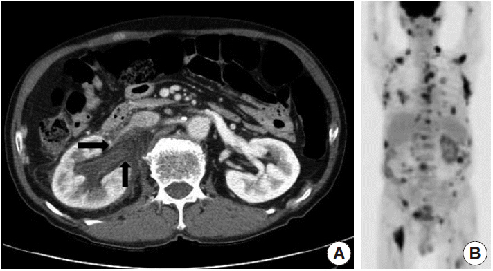
Fig. 1. (A) Contrast-enhanced axial computed tomography scan of the abdomen shows thickening with enhancement of the right renal pelvis wall with perinephric soft tissue infiltration (arrows) and hydroureteronephrosis. (B) Positron emission tomography computed tomography reveals hypermetabolic lesions in multiple neck, axillary, mediastinal, and pelvic lymph nodes as well as the chest, abdominal wall, left parotid gland, and right thigh.
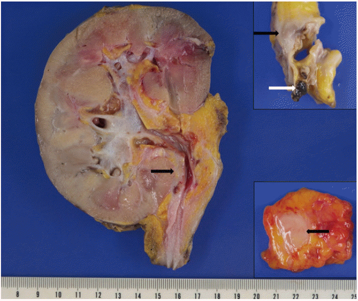
Fig. 2. Grossly, the specimen shows whitish yellow solid lesions along the renal pelvis and the ureter (black arrow), forming a concentric mass (black arrow, lower inset) compressing the ureter lumen. Renal artery surrounded by white solid lesions (black arrow, upper inset) shows atheromatous plaques and a thrombus (white arrow, upper inset).
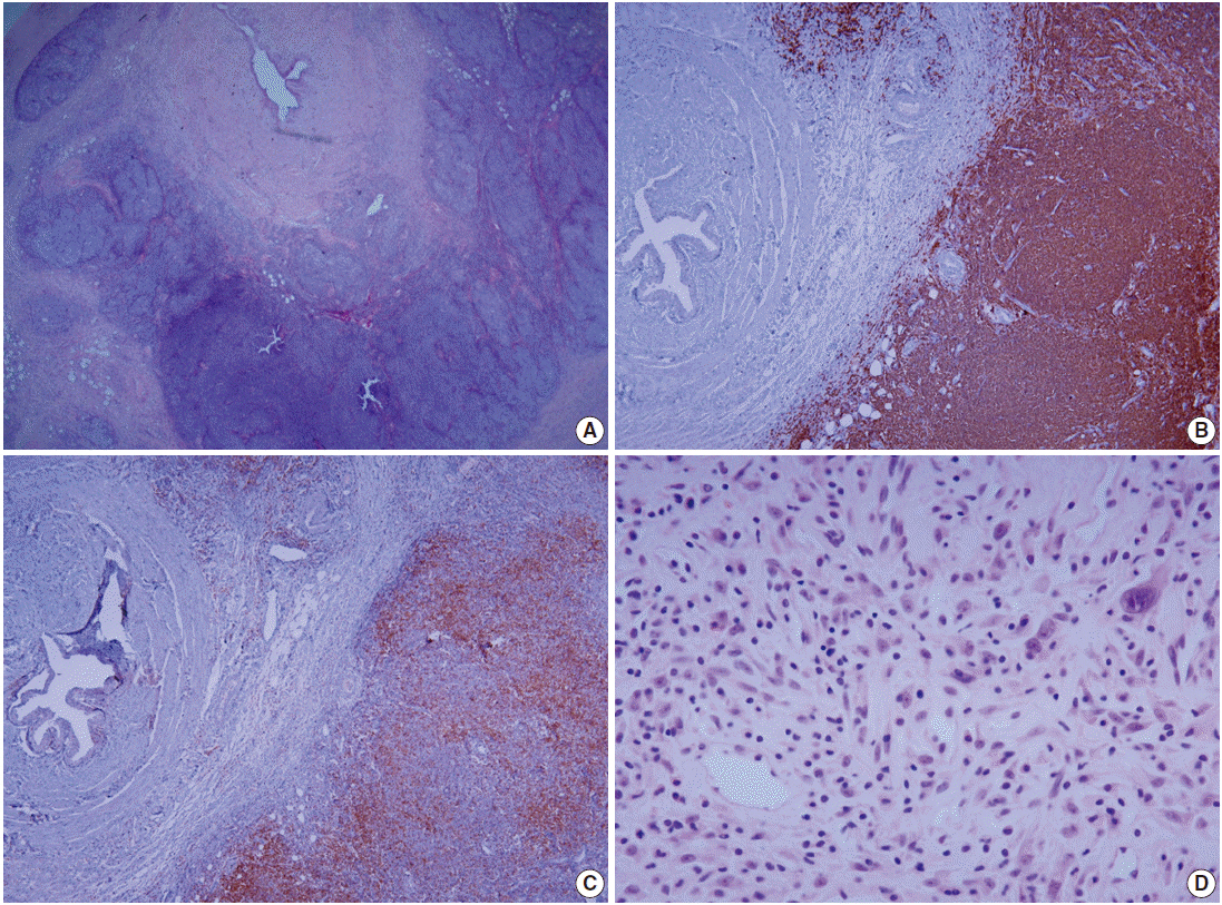
Fig. 3. Microscopic features of mucosa-associated lymphoid tissue lymphoma and adjacent chronic inflammation. (A) Tumor cells infiltrating the perimuscular layer of the ureter form lymphoid follicles with follicular colonization. Immunohistochemically, tumor cells are diffusely positive for CD20 (B) and accompanying T cells are mostly CD4 positive (C). (D) Peripelvic adipose tissue shows a mixed infiltrate of lymphoplasma cells, eosinophils, and histiocytes with fibroblastic proliferation.
Reference
-
1. Otsuki H, Ito K, Sato K, et al. Malignant lymphoma of mucosa-associated lymphoid tissue involving the renal pelvis and the entire ureter: a case report. Oncol Lett. 2013; 5:1625–8.
Article2. Thieblemont C, Bertoni F, Copie-Bergman C, Ferreri AJ, Ponzoni M. Chronic inflammation and extra-nodal marginal-zone lymphomas of MALT-type. Semin Cancer Biol. 2014; 24:33–42.
Article3. Eble JN, Sauter G, Epstein JI, Sesterhenn IA. World Health Organization classification of tumours: pathology and genetics tumors of the urinary system and male genital organs. Lyon: IARC Press;2004.4. Araki K, Kubota Y, Iijima Y, et al. Indolent behaviour of low-grade B-cell lymphoma of mucosa-associated lymphoid tissue involved in salivary glands, renal sinus and prostate. Scand J Urol Nephrol. 1998; 32:234–6.5. Qiu L, Unger PD, Dillon RW, Strauchen JA. Low-grade mucosa-associated lymphoid tissue lymphoma involving the kidney: report of 3 cases and review of the literature. Arch Pathol Lab Med. 2006; 130:86–9.
Article6. Mita K, Ohnishi Y, Edahiro T, Fujii T, Yamasaki A, Shimamoto F. Primary mucosa-associated lymphoid tissue lymphoma in the renal pelvis. Urol Int. 2002; 69:241–3.
Article7. Hara M, Satake M, Ogino H, et al. Primary ureteral mucosa-associated lymphoid tissue (MALT) lymphoma: pathological and radiological findings. Radiat Med. 2002; 20:41–4.8. Matsuda I, Zozumi M, Tsuchida YA, et al. Primary extranodal marginal zone lymphoma of mucosa-associated lymphoid tissue type with malakoplakia in the urinary bladder: a case report. Int J Clin Exp Pathol. 2014; 7:5280–4.9. Thieblemont C, Berger F, Dumontet C, et al. Mucosa-associated lymphoid tissue lymphoma is a disseminated disease in one third of 158 patients analyzed. Blood. 2000; 95:802–6.
Article10. Hansson GK, Robertson AK, SÖderberg-Nauclér C. Inflammation and atherosclerosis. Annu Rev Pathol. 2006; 1:297–329.
Article
- Full Text Links
-
- Actions
-
Cited
- CITED
-
- Close
- Share
-
- Similar articles
-
- A Case of Primary Pulmonary Extranodal Marginal Zone B-Cell Lymphoma of the MALT Type
- Role of Chemotherapy in Gastric Marginal Zone B-Cell Lymphoma of Mucosa-Associated Lymphoid Tissue (MALT) Type
- A Case of Primary Nodal Type Marginal Zone B-Cell Lymphoma of the Intra-Parotid Lymph Node Mistaken to Primary Benign Parotid Mass
- A Case of Mucosa-Associated Lymphoid Tissue Lymphoma in Nasopharynx and Thyroid Gland
- Endoscopic Findings of Gastric Extranodal Marginal Zone B-Cell Mucosa-Associated Lymphoid Tissue Lymphoma
