J Korean Ophthalmol Soc.
2015 Jul;56(7):998-1005. 10.3341/jkos.2015.56.7.998.
Assessment of Posterior Capsular Opacification of Korean Using Straylight and Glare Sensitivity Meter
- Affiliations
-
- 1Department of Ophthalmology and Visual Science, The Catholic University of Korea College of Medicine, Seoul, Korea. Sara514@catholic.ac.kr
- KMID: 2148798
- DOI: http://doi.org/10.3341/jkos.2015.56.7.998
Abstract
- PURPOSE
To evaluate posterior capsular opacity (PCO) using straylight and glare sensitivity meter and to compare availability of straylight and glare sensitivity with known methods for PCO evaluation.
METHODS
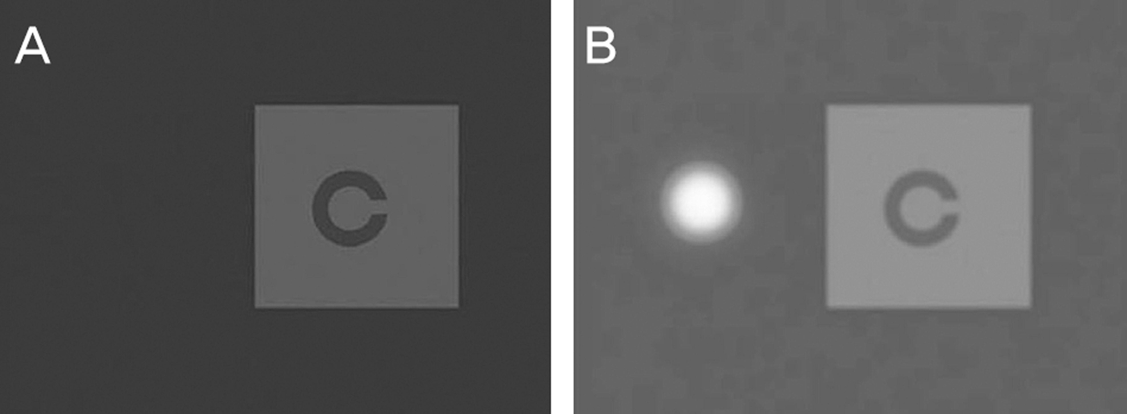
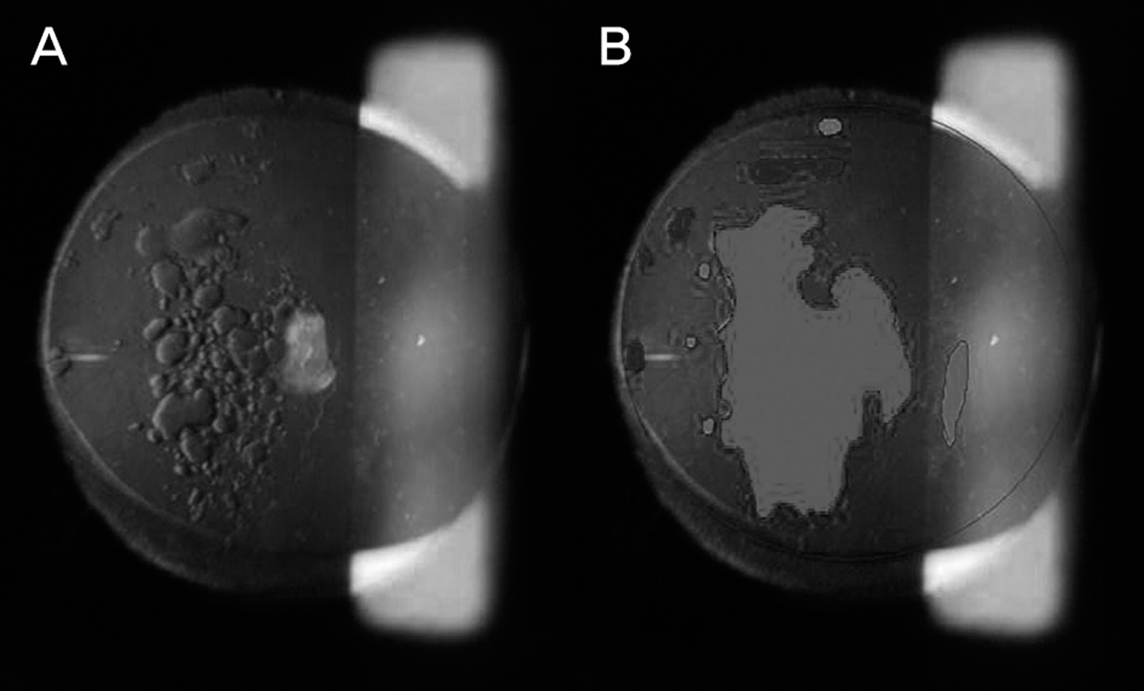
Thirty-six pseudophakic eyes with PCO were selected for this study. Best-corrected visual acuity (BCVA), straylight (C-quant, Oculus GmbH, Wetzlar, Germany) and glare sensitivity (Binoptometer, Oculus GmbH, Wetzlar, Germany) were measured before mydriasis. After mydriasis, PCO images were captured with a slit-lamp and analyzed using the Evaluation of Posterior Capsular Opacification (EPCO) program (EPCO software, University of Heidelberg, Heidelberg, Germany). The same measurements were taken after capsulotomy and compared with pre-capsulotomy data.
RESULTS
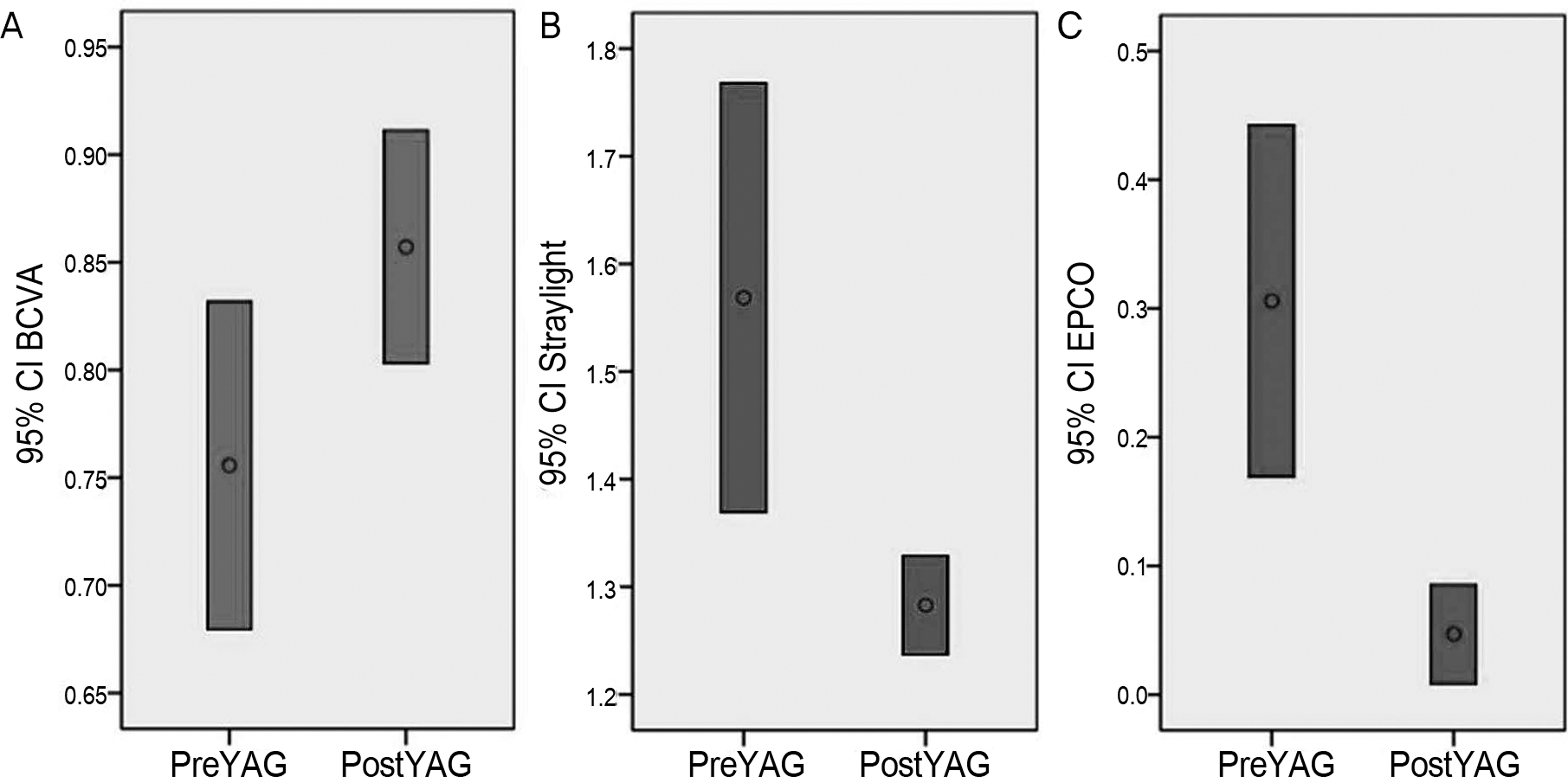
After capsulotomy, BCVA, EPCO score and straylight were improved with statistical significance (p < 0.05). Cases of PCO with mildly decreased visual acuity showed statistically significantly improved EPCO score and straylight (p < 0.05). Glare sensitivity did not show significant improvement but was statistically significantly correlated with straylight (p = 0.023, Rho = 0.732).
CONCLUSIONS
Straylight is an available measurement for evaluation of PCO. Glare sensitivity meter which correlates with straylight can be used as a supportive measurement.
MeSH Terms
Figure
Reference
-
References
1. Pandey SK, Apple DJ, Werner L. . Posterior capsule opacifica-tion: a review of the aetiopathogenesis, experimental and clinical studies and factors for prevention. Indian J Ophthalmol. 2004; 52:99–112.2. Cheng CY, Yen MY, Chen SJ. . Visual acuity and contrast sen-sitivity in different types of posterior capsule opacification. J Cataract Refract Surg. 2001; 27:1055–60.
Article3. van Bree MC, van den Berg TJ, Zijlmans BL. Posterior capsule opacification severity, assessed with straylight measurement, as main indicator of early visual function deterioration. Ophthalmology. 2013; 120:20–33.
Article4. van den Berg TJ. Introduction to retinal straylight. Netherlands Institute for Neuroscience. 2004; 1–11.5. Michael R, van Rijn LJ, van den Berg TJ. . Association of lens, opacities, intraocular straylight, contrast sensitivity and visual acuity in European drivers. Acta Ophthalmol. 2009; 87:666–71.
Article6. Aslam TM, Haider D, Murray IJ. Principles of disability glare measurement: an ophthalmological perspective. Acta Ophthalmol Scand. 2007; 85:354–60.
Article7. Kang MJ, Hwang HB, Chung SK. Effect of glistening-free intra-ocular lens on intraocular straylight. J Korean Ophthalmol Soc. 2014; 55:1001–6.
Article8. Lee SY, Oh JH. Straylight in normal and cataractous eyes of Koreans. J Korean Ophthalmol Soc. 2011; 52:182–9.
Article9. Hiraoka T, Okamoto C, Ishii Y. . Mesopic contrast sensitivity and ocular higher-order aberrations after overnight orthokeratology. Am J Ophthalmol. 2008; 145:645–55.
Article10. Puell MC, Palomo C, Sánchez-Ramos C, Villena C. Mesopic con-trast sensitivity in the presence or absence of glare in a large driver population. Graefes Arch Clin Exp Ophthalmol. 2004; 242:755–61.
Article11. Findl O, Buehl W, Menapace R. . Comparison of 4 methods for quantifying posterior capsule opacification. J Cataract Refract Surg. 2003; 29:106–11.
Article12. Tetz MR, Auffarth GU, Sperker M. . Photographic image anal-ysis system of posterior capsule opacification. J Cataract Refract Surg. 1997; 23:1515–20.
Article13. Van Den Berg TJ, Van Rijn LJ, Michael R. . Straylight effects with aging and lens extraction. Am J Ophthalmol. 2007; 144:358–63.
Article14. Langeslag MJ, van der Mooren M, Beiko GH, Piers PA. Impact of intraocular lens material and design on light scatter: in vitro study. J Cataract Refract Surg. 2014; 40:2120–7.
Article15. Hirnschall N, Crnej A, Gangwani V, Findl O. Comparison of meth-ods to quantify posterior capsule opacification using forward and backward light scattering. J Cataract Refract Surg. 2014; 40:728–35.
Article16. Montenegro GA, Marvan P, Dexl A. . Posterior capsule opaci-fication assessment and factors that influence visual quality after posterior capsulotomy. Am J Ophthalmol. 2010; 150:248–53.
Article17. Franssen L, Tabernero J, Coppens JE, van den Berg TJ. Pupil size and retinal straylight in the normal eye. Invest Ophthalmol Vis Sci. 2007; 48:2375–82.
Article18. van der Meulen IJ, Gjertsen J, Kruijt B. . Straylight measure-ments as an indication for cataract surgery. J Cataract Refract Surg. 2012; 38:840–8.
Article
- Full Text Links
- Actions
-
Cited
- CITED
-
- Close
- Share
- Similar articles
-
- A Case of Liquefied Posterior Capsular Opacification
- Straylight in Normal and Cataractous Eyes of Koreans
- The Effect of Two Different Opening Patterns of Neodymium:YAG Laser Posterior Capsulotomy on Visual Function
- The Effect of Anterior Capsulotomy Size and Lens Epithelial cells Removal on the Posterior Capsular Opacification
- The Rate of Posterior Capsular Opacification with Three Different Sizes of Capsular Intraocular Lenses


