Unexpected Pathologic Diagnosis of the Mitral Valvular Mass
- Affiliations
-
- 1Division of Cardiology, Department of Internal Medicine, Cheil General Hospital, Dankook University College of Medicine, Seoul, Korea.
- 2Division of Cardiology, Department of Internal Medicine, Korea University Anam Hospital, Korea University College of Medicine, Seoul, Korea. smparkmd@korea.ac.kr
- 3Department of Radiology, Korea University Anam Hospital, Korea University College of Medicine, Seoul, Korea.
- 4Department of Thoracic Surgery, Korea University Anam Hospital, Korea University College of Medicine, Seoul, Korea.
- KMID: 2144460
- DOI: http://doi.org/10.4250/jcu.2015.23.4.271
Abstract
- A 59-year-old man with multifocal cerebral infarction was found to have the large obstructive mitral valvular mass. Although benign tumor was under suspicion before surgery, he was finally diagnosed as chronic infective endocarditis by microscopic evaluation. The precise diagnosis and the proper management of a cardiac mass are very important since even the benign tumor may cause fatal complications. However, primary cardiac mass has the broad spectrum from pseudo-tumor to malignancy and the differential diagnosis using non-invasive methods is not easy even with the currently available imaging techniques.
Figure
-
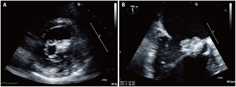
Fig. 1 Transthoracic (A) and transesophageal (B) echocardiographic images of the mitral valvular mass. The mass was located on lateral mitral commissure, which made severe mitral stenosis. The surface of mass had several irregular with oscillating strands and tags.
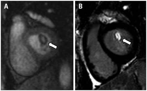
Fig. 2 Early perfusion image (A) and late gadolinium enhance image (B) of the mitral valve (arrow). At early perfusion image, the mass showed low signal intensity whereas high signal intensity was detected on late gadolinium enhance image.

Fig. 3 Gross image of the mitral valve apparatus shows well demarcated mass with greyish surface and filthy, amorphous oscillating structures on it in atrial side (A), but grossly normal valvular surface in ventricular side (B).
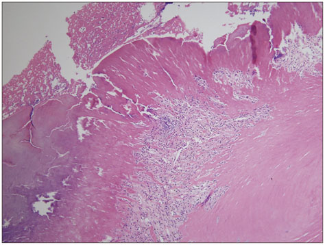
Fig. 4 Microscopic finding (hematoxylin and eosin, × 100) of the mitral valvular mass were composed of extensively thickened valvular leaflets with organizing thrombus. On valvular structure, plenty of lymphocytes and plasma cells on atrial surface and myxoid degeneration in ventricular side were presented.
Reference
-
1. Motwani M, Kidambi A, Herzog BA, Uddin A, Greenwood JP, Plein S. MR imaging of cardiac tumors and masses: a review of methods and clinical applications. Radiology. 2013; 268:26–43.2. Shapiro LM. Cardiac tumours: diagnosis and management. Heart. 2001; 85:218–222.3. Joffe II, Jacobs LE, Owen AN, Ioli A, Kotler MN. Noninfective valvular masses: review of the literature with emphasis on imaging techniques and management. Am Heart J. 1996; 131:1175–1183.4. Dursun M, Yılmaz S, Yılmaz E, Yılmaz R, Onur İ, Oflaz H, Dindar A. The utility of cardiac MRI in diagnosis of infective endocarditis: preliminary results. Diagn Interv Radiol. 2015; 21:28–33.5. Habib G, Lancellotti P, Antunes MJ, Bongiorni MG, Casalta JP, Del Zotti F, Dulgheru R, El Khoury G, Erba PA, Iung B, Miro JM, Mulder BJ, Plonska-Gosciniak E, Price S, Roos-Hesselink J, Snygg-Martin U, Thuny F, Tornos Mas P, Vilacosta I, Zamorano JL, Erol Ç, Nihoyannopoulos P, Aboyans V, Agewall S, Athanassopoulos G, Aytekin S, Benzer W, Bueno H, Broekhuizen L, Carerj S, Cosyns B, De Backer J, De Bonis M, Dimopoulos K, Donal E, Drexel H, Flachskampf FA, Hall R, Halvorsen S, Hoen B, Kirchhof P, Lainscak M, Leite-Moreira AF, Lip GY, Mestres CA, Piepoli MF, Punjabi PP, Rapezzi C, Rosenhek R, Siebens K, Tamargo J, Walker DM. Authors/Task Force Members. Document Reviewers. 2015 ESC Guidelines for the management of infective endocarditis: The Task Force for the Management of Infective Endocarditis of the European Society of Cardiology (ESC)Endorsed by: European Association for Cardio-Thoracic Surgery (EACTS), the European Association of Nuclear Medicine (EANM). Eur Heart J. 2015; 36:3075–3128.6. Thiene G, Basso C. Pathology and pathogenesis of infective endocarditis in native heart valves. Cardiovasc Pathol. 2006; 15:256–263.
- Full Text Links
-
- Actions
-
Cited
- CITED
-
- Close
- Share
-
- Similar articles
-
- Relation between Left Atrial Size and Atrial Fibrillation
- Familial mitral valve prolapse in a Maltese dog family
- The Relationship of Mitral Valve Area Measured by 2-Dimensional Echocardiography with the M-Mode Measurements in Mitral Valvular Stenosis
- Two women presenting aborted sudden cardiac arrest as the first event of mitral valve disease
- Echocardiographic Study on the Mitral Valvular Heart Diseases
