Two women presenting aborted sudden cardiac arrest as the first event of mitral valve disease
- Affiliations
-
- 1Department of Critical Care Medicine, Samsung Medical Center, Sungkyunkwan University School of Medicine, Seoul, Korea.
- 2Division of Cardiology, Department of Internal Medicine, Korea University Anam Hospital, Korea University College of Medicine, Seoul, Korea. smparkmd@korea.ac.kr
- KMID: 2467310
- DOI: http://doi.org/10.4266/acc.2017.00570
Abstract
- Mitral valve prolapse (MVP) is a relatively common valvular heart disease and is known to have a benign course. However, a certain subtype of MVP has a pathologic prognosis and can be accompanied by malignant cardiac arrhythmia causing sudden cardiac arrest, which can be characterized by bileaflet mitral valvular thickening and prolapse and frequent premature ventricular ectopic activity upon electrocardiography. Herein, we present two patients with bileaflet mitral prolapse who survived aborted sudden cardiac arrest. These cases show a precise MVP diagnosis that may prevent a devastating life event with the unique MVP subtype.
MeSH Terms
Figure
-
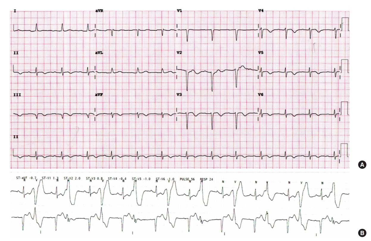
Figure 1. Electrocardiogram of case 1 presenting T wave inversion of inferior leads (A) and ventricular bigeminy (B).

Figure 2. Echocardiographic imaging of case 1. Two-dimensional image of parasternal long axis view (A) and apical two-chamber view (B) show bileaflet mitral valvular prolapse (red arrow) and backward displacement of the mitral annular ring (white arrow). Apical two-chamber view additively shows a hypertrophied papillary muscle (arrowhead). (C) Color Doppler image of the apical fourchamber view shows moderate mitral regurgitation (arrowhead).
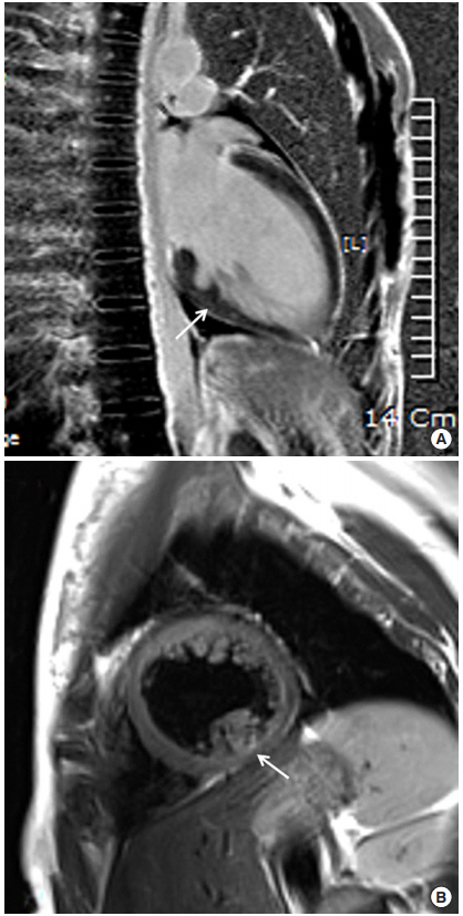
Figure 3. Cardiac magnetic resonance imaging of case 1 shows a thinned inferobasal left ventricular wall (A, arrow) and inhomogenous signal intensity by late gadolinium enhancement suggesting damaged myocardium (B, arrow).
Reference
-
1. Batra AS, Balaji S. Prevalence and spectrum diseases predisposing to sudden cardiac death: are they the same for both the athlete and the nonathlete? Pediatr Cardiol. 2012; 33:379–86.
Article2. Angella FR, Lewis JF. Mitral valve prolapse: gender differences in evaluation and management. Cardiol Rev. 1999; 7:161–8.3. Kligfield P, Levy D, Devereux RB, Savage DD. Arrhythmias and sudden death in mitral valve prolapse. Am Heart J. 1987; 113:1298–307.
Article4. Nishimura RA, McGoon MD, Shub C, Miller FA Jr, Ilstrup DM, Tajik AJ. Echocardiographically documented mitral-valve prolapse: long-term follow-up of 237 patients. N Engl J Med. 1985; 313:1305–9.5. Sriram CS, Syed FF, Ferguson ME, Johnson JN, Enriquez-Sarano M, Cetta F, et al. Malignant bileaflet mitral valve prolapse syndrome in patients with otherwise idiopathic out-of-hospital cardiac arrest. J Am Coll Cardiol. 2013; 62:222–30.
Article6. Basso C, Perazzolo Marra M, Rizzo S, De Lazzari M, Giorgi B, Cipriani A, et al. Arrhythmic mitral valve prolapse and sudden cardiac death. Circulation. 2015; 132:556–66.
Article7. Perazzolo Marra M, Basso C, De Lazzari M, Rizzo S, Cipriani A, Giorgi B, et al. Morphofunctional abnormalities of mitral annulus and arrhythmic mitral valve prolapse. Circ Cardiovasc Imaging. 2016; 9:e005030.
Article8. Anders S, Said S, Schulz F, Püschel K. Mitral valve prolapse syndrome as cause of sudden death in young adults. Forensic Sci Int. 2007; 171:127–30.
Article
- Full Text Links
-
- Actions
-
Cited
- CITED
-
- Close
- Share
-
- Similar articles
-
- Medullary Infarction Presenting as Sudden Cardiac Arrest: Report of Two Cases and Review of the Literature
- Free-Floating Left Atrial Thrombus with Recurrent cerebral Embolic Event Associated Mitral Stenosis
- Mitral “Hole-in-one Embolus†Detected by Transesophageal Echocardiography During Cardiopulmonary Resuscitation: A Cases Report
- A Case of Aborted Sudden Cardiac Death during Exercise Associated with an Anomalous Origin of Right Coronary Artery
- Usefulness of Pressure Half Time by Pulse Doppler Ultrasound in Evaluation of the Severity of Mitral Stenosis
