Clinical Application of Whole-body MRI
- Affiliations
-
- 1Department of Radiology, Sungkyunkwan University School of Medicine, Korea. jhkate@skku.edu
- KMID: 2137746
- DOI: http://doi.org/10.5124/jkma.2008.51.11.1034
Abstract
- Whole body magnetic resonance imaging (WB MRI) has become feasible and enables fast scan throughout the body. This technique is based on a real-time gradient-echo imaging and sliding table platform (rolling table concept, which eliminates time-consuming repositioning of patients and surface coils). MRI scanners of the latest generation use high field MRI units of >1.5 Tesla (T), and are reported to have upgraded capabilities in terms of temporal and spatial resolution due to improved signal-to-noise ratios (SNRs) under high magnetic-field strength conditions. The diagnostic accuracy of the whole-body staging strategies of PET/CT and MRI are established. As a start of tumor staging through whole body imaging, PET/CT showed superior performances in T and N staging than WB MRI using 1.5T MR system. But, both imaging procedures showed a similar performance in detecting distant metastases. In a recent report on staging of non-small cell lung cancer (NSCLC), whole body MR imaging proved to be effective as much as PET/CT in T, N, and M staging. In addition, there were organs of strength for each modality in the detection of metastasis. Therefore, whole-body MRI/ PET would be suggested as a future diagnostic procedure of choice for the whole-body imaging with synergistic effects and reduced radiation exposure.
Keyword
MeSH Terms
Figure
-

Figure 1 Concordant detection of metastasis in a 48-year-old woman with adenocarcinoma in the right lung. (A) Max-imum intensity projection PET image shows a hyper-metabolic mass (arrow) in the left temporal lobe of the brain and a focal hot-uptake lesion (double- arrow) in the left iliac bone, representing brain and bone metastases. (B) Enhanced T1-weighted turbo field-echo whole-body MR image shows a rim-enhancing mass (arrow) in the left temporal lobe of the brain and a focal enhancing lesion (double-arrow) in the left iliac bone, representing brain and bone metastases. Note another smaller metastatic nodule (arrowhead) in the left thalamus of the brain, which was not covered in the field-of-view of the PET/CT.
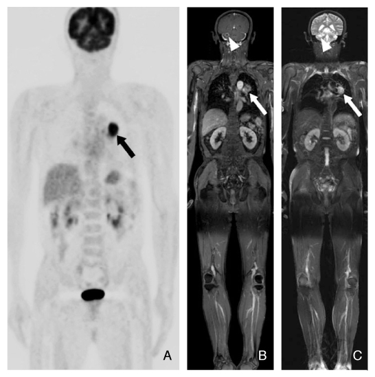
Figure 2 Images of brain metastasis in a 62-year-old man with NSCLC of the lung detected at whole-body MR imaging, but not at PET/CT. (A) Maximum intensity projection PET image shows a primary lung cancer (arrow) in the left upper lobe, but not metastatic nodule in the brain. (B) Enhanced T1-weighted turbo field -echo whole-body MR image shows a spiculated enhancing primary lung cancer in the left upper lobe (arrow) and a rim-enhancing metastatic nodule (arrowhead) in right cerebella hemisphere in the brain. (C) T2-weighted turbo spin-echo whole-body MR image shows a focal high signal intensity lesion (arrowhead) in the corresponding area of the right cerebella hemisphere. Note primary mass in left upper lobe (arrow). Increase in size on follow-up MR images confirms a brain metastasis.
Reference
-
1. Horvath LJ, Burtness BA, McCarthy S, Johnson KM. Total-body echo-planar MR imaging in the staging of breast cancer: comparison with conventional methods-early experience. Radiology. 1999. 211:119–128.
Article2. Walker R, Kessar P, Blanchard R, Dimasi M, Harper K, DeCarvalho V, Yucel EK, Patriquin L, Eustace S. Turbo STIR magnetic resonance imaging as a whole-body sc-reening tool for metastases in patients with breast car-cinoma: preliminary clinical experience. J Magn Re-son Imagin. 2000. 11:343–350.
Article3. Lauenstein TC, Goehde SC, Herborn CU, Goyen M, Ober-hoff C, Debatin JF, Ruehm SG, Barkhausen J. Whole-body MR imaging: evaluation of patients for metastases. Radiology. 2004. 233:139–148.
Article4. Yi CA, Shin KM, Lee KS, Kim BT, Kim H, Kwon OJ, Choi JY, Chung MJ. Non-small cell lung cancer staging: efficacy comparison of integrated PET/CT versus 3.0-T whole-body MR imaging. Radiology. 2008. 248:632–642.
Article5. Barkhausen J, Quick HH, Lauenstein T, Goyen M, Ruehm SG, Laub G, Debatin JF, Ladd ME. Whole-body MR imaging in 30 seconds with real-time true FISP and a continuously rolling table platform: feasibility study. Radiology. 2001. 220:252–256.
Article6. Thomson V, Pialat JB, Gay F, Coulon A, Voloch A, Granier A, Guerin JC, Viallon M, Berthezene Y. Whole-body MRI for metastases screening: a preliminary study using 3D VIBE sequences with automatic subtraction between noncontrast and contrast enhanced images. American journal of clinical oncology. 2008. 31:285–292.
Article7. Darge K, Jaramillo D, Siegel MJ. Whole-body MRI in children: Current status and future applications. Eur-opean journal of radiology. 2008.
Article8. Daldrup-Link HE, Franzius C, Link TM, Laukamp D, Sciuk J, Jurgens H, Schober O, Rummeny EJ. Whole-body MR imaging for detection of bone metastases in children and young adults: comparison with skeletal scintigraphy and FDG PET. Ajr. 2001. 177:229–236.
Article9. Mazumdar A, Siegel MJ, Narra V, Luchtman-Jones L. Whole-body fast inversion recovery MR imaging of small cell neoplasms in pediatric patients: a pilot study. Ajr. 2002. 179:1261–1266.
Article10. Laffan EE, O'Connor R, Ryan SP, Donoghue VB. Whole-body magnetic resonance imaging: a useful additional sequence in paediatric imaging. Pediatric radiology. 2004. 34:472–480.
Article11. Goo HW, Choi SH, Ghim T, Moon HN, Seo JJ. Whole-body MRI of paediatric malignant tumours: comparison with conventional oncological imaging methods. Pediatric radiology. 2005. 35:766–773.
Article12. Lauenstein TC, Semelka RC. Emerging techniques: whole-body screening and staging with MRI. J Magn Reson Imaging. 2006. 24:489–498.
Article13. Althoff CE, Appel H, Rudwaleit M, Sieper J, Eshed I, Hamm B, Hermann KG. Whole-body MRI as a new screening tool for detecting axial and peripheral mani-festations of spondyloarthritis. Annals of the rheumatic diseases. 2007. 66:983–985.
Article14. Ladd SC, Debatin JF, Stang A, Bromen K, Moebus S, Nuefer M, Gizewski E, Wanke I, Doerfler A, Ladd ME, Benemann J, Erbel R, Forsting M, Schmermund A, Jockel KH. Whole-body MR vascular screening detects unsuspected concomitant vascular disease in coronary heart disease patients. European radiology. 2007. 17:1035–1045.
Article
- Full Text Links
-
- Actions
-
Cited
- CITED
-
- Close
- Share
-
- Similar articles
-
- Whole-Body MRI in Children: Current Imaging Techniques and Clinical Applications
- Principle, Development, and Application of Electrical Conductivity Mapping Using Magnetic Resonance Imaging
- Adnexal Masses: Clinical Application of Multiparametric MR Imaging & O-RADS MRI
- PET/MRI: Technical Challenges and Recent Advances
- Clinical Application and Limitations of Myeloma Response Assessment and Diagnosis System (MY-RADS)
