J Korean Assoc Oral Maxillofac Surg.
2012 Dec;38(6):332-336. 10.5125/jkaoms.2012.38.6.332.
A comparison of fixation methods using three-dimensional finite element analysis following anterior segmental osteotomy
- Affiliations
-
- 1Department of Oral and Maxillofacial Surgery, The Catholic University of Korea, St. Paul's Hospital, Seoul, Korea.
- 2Department of Oral and Maxillofacial Surgery, College of Medicine, The Catholic University of Korea, Seoul, Korea. jupark@catholic.ac.kr
- 3Department of Mechanical Engineering, Myongji University, Seoul, Korea.
- KMID: 2136969
- DOI: http://doi.org/10.5125/jkaoms.2012.38.6.332
Abstract
OBJECTIVES
This study sought to evaluate fixation methods and determine the best method for the postoperative stabilization of maxillary osteotomy. For our analysis we performed a three-dimensional finite element analysis of stress distribution on the plate, screw, and surrounding bone, as well as displacement onto the plate.
MATERIALS AND METHODS
We generated a model using synthetic skull scan data; an initital surface model was changed to a solid model using software. Modified anterior segmental osteotomy (using Park's method) was made using the program, and four different types of fixation methods were used. An anterior load of 100 N was applied on the palatal surface of two central incisors.
RESULTS
The Type 1 (L-shaped) fixation method gave stresses of 187.8 MPa at the plate, 45.8 MPa at the screw, and 15.4 MPa at the bone around the plate. The Type 2 (I-shaped) fixation method gave stresses of 186.6 MPa at the plate, 75.7 MPa at the screw, and 13.8 MPa at the bone around the plate. The Type 3 (inverted L-shaped) fixation method gave stresses of 28.6 MPa at the plate, 29.9 MPa at the screw, and 15.3 MPa at the bone around the plate. The Type 4 (I-shaped) fixation method gave stresses of 34.8 MPa at the plate, 36.9 MPa at the screw, and 14.9 MPa at the bone around the plate. The deflection of the plates for the four fixation methods was 0.014 mm, 0.022 mm, 0.017 mm, and 0.018 mm, respectively.
CONCLUSION
The Type 3 (inverted L-shaped) fixation method offers more stability than the other fixation methods. We therefore recommend this method for the postoperative stabilization of maxillary osteotomy.
Figure
-
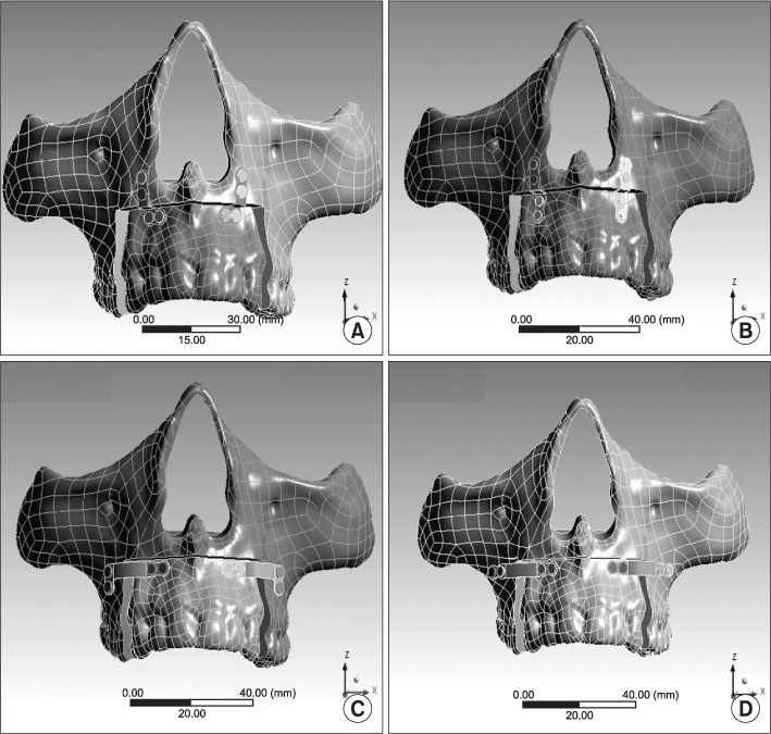
Fig. 1 Four types of anterior segmental osteotomy model fixed with different pattern of plates and screws. A. Type 1. B. Type 2. C. Type 3. D. Type 4.
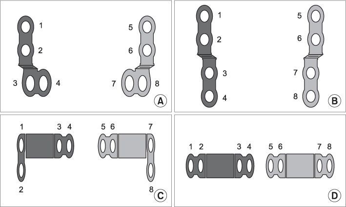
Fig. 2 The screw holes were numbered by their positions. A. Type 1. B. Type 2. C. Type 3. D. Type 4.
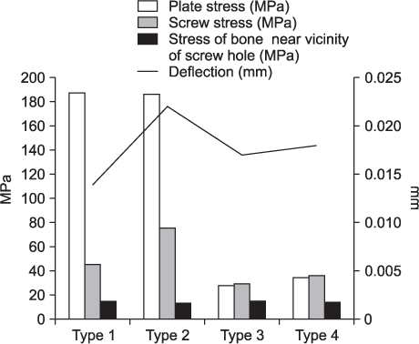
Fig. 3 Comparison of von Mises stress of plates, screws, bone near screw holes, and deflection of plates of 4 different types.
Reference
-
1. Bell WH, Proffit WR, White RP. Surgical correction of dentofacial deformities. 1980. Philadelphia: Saunders;1015.2. Chu YM, Po-Hsun Chen R, Morris DE, Wen-Ching Ko E, Chen YR. Surgical approach to the patient with bimaxillary protrusion. Clin Plast Surg. 2007. 34:535–546.
Article3. Baek SH, Kim BH. Determinants of successful treatment of bimaxillary protrusion: orthodontic treatment versus anterior segmental osteotomy. J Craniofac Surg. 2005. 16:234–246.
Article4. Jacobs JD, Bell WH. Combined surgical and orthodontic treatment of bimaxillary protrusion. Am J Orthod. 1983. 83:321–333.
Article5. Rosenquist B. Anterior segmental maxillary osteotomy. A 24-month follow-up. Int J Oral Maxillofac Surg. 1993. 22:210–213.6. Park JU. Orthognathic surgery. 2003. 1st ed. Seoul: Koonja Publishing;267.7. Park JU, Hwang YS. Evaluation of the soft and hard tissue changes after anterior segmental osteotomy on the maxilla and mandible. J Oral Maxillofac Surg. 2008. 66:98–103.
Article8. Ataç MS, Erkmen E, Yücel E, Kurt A. Comparison of biomechanical behaviour of maxilla following Le Fort I osteotomy with 2- versus 4-plate fixation using 3D-FEA. Part 1: advancement surgery. Int J Oral Maxillofac Surg. 2008. 37:1117–1124.
Article9. Poiate IA, Vasconcellos AB, Andueza A, Pola IR, Poiate E Jr. Three dimensional finite element analyses of oral structures by computerized tomography. J Biosci Bioeng. 2008. 106:606–609.
Article10. Tanne K, Miyasaka J, Yamagata Y, Sachdeva R, Tsutsumi S, Sakuda M. Three-dimensional model of the human craniofacial skeleton: method and preliminary results using finite element analysis. J Biomed Eng. 1988. 10:246–252.
Article11. Tanne K, Sakuda M, Burstone CJ. Three-dimensional finite element analysis for stress in the periodontal tissue by orthodontic forces. Am J Orthod Dentofacial Orthop. 1987. 92:499–505.
Article12. Proffit WR, Sarver DM. Proffit WR, Fields HW, Sarver DM, editors. Combined surgical and orthodontic treatment. Contemporary orthodontics. 2007. St. Louis: Elsevier Inc.;686.13. Ziccardi VB, Wilk R. Fonseca RJ, editor. Anterior and posterior maxillary segmental osteotomies. Oral and maxillofacial surgery. 2000. Philadelphia: Saunders;249.14. Korkmaz HH. Evaluation of different miniplates in fixation of fractured human mandible with the finite element method. Oral Surg Oral Med Oral Pathol Oral Radiol Endod. 2007. 103:e1–e13.
Article15. Gunaseelan R, Anantanarayanan P, Veerabahu M, Vikraman B, Sripal R. Intraoperative and perioperative complications in anterior maxillary osteotomy: a retrospective evaluation of 103 patients. J Oral Maxillofac Surg. 2009. 67:1269–1273.
Article16. Vollmer D, Meyer U, Joos U, Vègh A, Piffko J. Experimental and finite element study of a human mandible. J Craniomaxillofac Surg. 2000. 28:91–96.
Article17. Ataç MS, Erkmen E, Yücel E, Kurt A. Comparison of biomechanical behaviour of maxilla following Le Fort I osteotomy with 2- versus 4-plate fixation using 3D-FEA Part 2: impaction surgery. Int J Oral Maxillofac Surg. 2009. 38:58–63.
Article18. Erkmen E, Ataç MS, Yücel E, Kurt A. Comparison of biomechanical behaviour of maxilla following Le Fort I osteotomy with 2- versus 4-plate fixation using 3D-FEA: part 3: inferior and anterior repositioning surgery. Int J Oral Maxillofac Surg. 2009. 38:173–179.
Article19. Alberts LR, Phillips KO, Tu HK, Stinson WW, Friedman A. A biologic model for assessment of osseous strain patterns and plating systems in the human maxilla. J Oral Maxillofac Surg. 2003. 61:79–88.
Article20. Armstrong JE, Lapointe HJ, Hogg NJ, Kwok AD. Preliminary investigation of the biomechanics of internal fixation of sagittal split osteotomies with miniplates using a newly designed in vitro testing model. J Oral Maxillofac Surg. 2001. 59:191–195.
Article21. Song HC, Throckmorton GS, Ellis E 3rd, Sinn DP. Functional and morphologic alterations after anterior or inferior repositioning of the maxilla. J Oral Maxillofac Surg. 1997. 55:41–49.
Article
- Full Text Links
-
- Actions
-
Cited
- CITED
-
- Close
- Share
-
- Similar articles
-
- Finite element analysis of anterior maxillary segmental distraction osteogenesis using asymmetric distractors in patients with unilateral cleft lip and palate
- Finite Element Model of A-P Instrimentation in Thoracolumbar Burst Fracture
- A three-dimensional Finite element analysis for initial stress of maxillary incisors during activation of upper utility arch wire
- Three-dimensional finite element analysis of the stress distribution and displacement in different fixation methods of bilateral sagittal split ramus osteotomy
- Three dimensional finite element analysis of mandibular stresses of complete denture occlusion
