Chronic maxillary sinusitis and diabetes related maxillary osteonecrosis: a case report
- Affiliations
-
- 1Department of Oral and Maxillofacial Surgery, Kyung Hee University School of Dentistry, Seoul, Korea. yongdae.kwon@gmail.com
- KMID: 2133181
- DOI: http://doi.org/10.5125/jkaoms.2015.41.6.332
Abstract
- Dental infections and maxillary sinusitis are the main causes of osteomyelitis. Osteomyelitis can occur in all age groups, and is more frequently found in the lower jaw than in the upper jaw. Systemic conditions that can alter the patient's resistance to infection including diabetes mellitus, anemia, and autoimmune disorders are predisposing factors for osteomyelitis. We report a case of uncommon broad maxillary osteonecrosis precipitated by uncontrolled type 2 diabetes mellitus and chronic maxillary sinusitis in a female patient in her seventies with no history of bisphosphonate or radiation treatment.
MeSH Terms
Figure
-
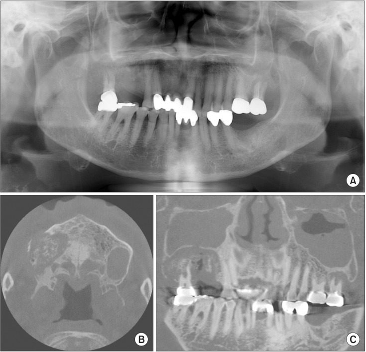
Fig. 1 Radiographic exams at the first visit show relatively well defined bone destructive lesion (sequestrum) on right maxillary molar area with elevation of sinus floor. A. Panoramic radiograph. B, C. Cone-beam computed tomography.
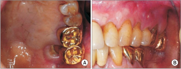
Fig. 2 Intra-oral photographs show oro-antral fistula (palate; A) and bony exposure (buccal; B) on left maxilla after 14 months since the first visit.
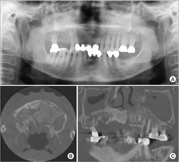
Fig. 3 Radiographic exams show severe bony destructive lesions on both maxilla and mucosal thickening on both maxillary sinuses after 14 months since the first visit. A. Panoramic radiograph. B, C. Cone-beam computed tomography.
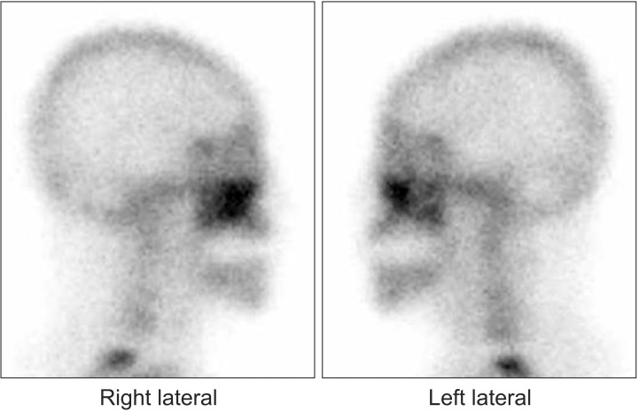
Fig. 4 Bone scan; active bony lesions in both maxilla.
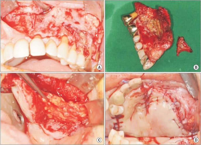
Fig. 5 Clinical photographs were taken during operation (A) extensive necrotic destruction of the both maxilla (B) excised specimen of left maxilla and palate (C) reconstruction with buccal fat pad flap (D) primary wound closure.
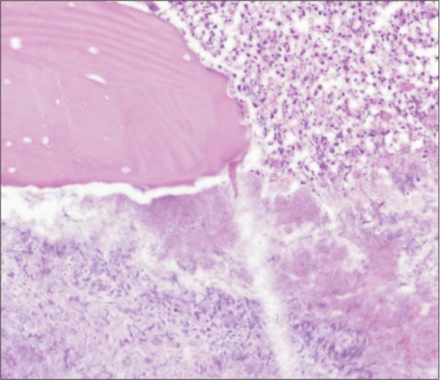
Fig. 6 Histopathologically, bony tissues show bony necrosis, marrow fibrosis and numerous sulfur granules with acute and chronic inflammatory cell infiltration (H&E staining, ×200).
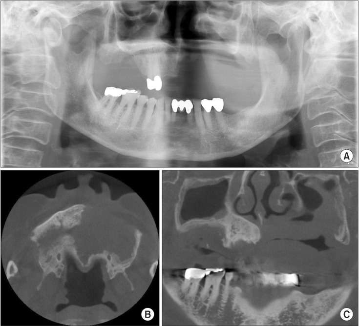
Fig. 7 Postoperative radiographic exams. A. Panoramic radiograph. B, C. Cone-beam computed tomography.

Fig. 8 A. Intra-oral photographic with removable partial denture. B. No signs of recurrence after 13 months of follow-up.
Reference
-
1. Macbeth R. Osteomyelitis of the maxilla. J Laryngol Otol. 1952; 66:18–28. PMID: 14898169.
Article2. Indrizzi E, Terenzi V, Renzi G, Bonamini M, Bartolazzi A, Fini G. The rare condition of maxillary osteomyelitis. J Craniofac Surg. 2005; 16:861–864. PMID: 16192871.
Article3. Hudson JW. Osteomyelitis of the jaws: a 50-year perspective. J Oral Maxillofac Surg. 1993; 51:1294–1301. PMID: 8229407.
Article4. Schenck HP. Osteomyelitis of the frontal bone. Ann Otol Rhinol Laryngol. 1959; 68:336–345. PMID: 13661805.
Article5. Prasad KC, Prasad SC, Mouli N, Agarwal S. Osteomyelitis in the head and neck. Acta Otolaryngol. 2007; 127:194–205. PMID: 17364352.6. Koorbusch GF, Fotos P, Goll KT. Retrospective assessment of osteomyelitis. Etiology, demographics, risk factors, and management in 35 cases. Oral Surg Oral Med Oral Pathol. 1992; 74:149–154. PMID: 1508521.7. Adekeye EO, Cornah J. Osteomyelitis of the jaws: a review of 141 cases. Br J Oral Maxillofac Surg. 1985; 23:24–35. PMID: 3156622.8. Borle RM, Borle SR. Osteomyelitis of the zygomatic bone: a case report. J Oral Maxillofac Surg. 1992; 50:296–298. PMID: 1542073.
Article9. Reid IR. Osteonecrosis of the jaw: who gets it, and why? Bone. 2009; 44:4–10. PMID: 18948230.
- Full Text Links
-
- Actions
-
Cited
- CITED
-
- Close
- Share
-
- Similar articles
-
- Maxillary sinusitis as a complication of oral bisphosphonate related osteonecrosis of the jaw: A case report
- Delayed Occurrence of Maxillary Sinusitis after Simultaneous Maxillary Sinus Augmentation and Implant: A Case Report and Literature Review
- A clinical study on maxillary sinusitis in children with respiratory allergic disease
- A bacteriological study in Caldwell-Luc's operation of chronic maxillary sinusitis
- Organized Hematoma Presenting with Periorbital Swelling: A Case Report and Review of Literatures
