Five-year retrospective radiographic follow-up study of dental implants with sandblasting with large grit, and acid etching-treated surfaces
- Affiliations
-
- 1Department of Oral and Maxillofacial Surgery, Chungnam National University School of Medicine, Daejeon, Korea. kjjomfs@cnu.ac.kr
- 2Department of Oral and Maxillofacial Surgery, Chungbuk National University School of Medicine, Cheongju, Korea.
- KMID: 2133178
- DOI: http://doi.org/10.5125/jkaoms.2015.41.6.317
Abstract
OBJECTIVES
The purpose of this study is to evaluate five-year radiographic follow-up results of the Korean sandblasting with large grit, and acid etching (SLA)-treated implant system.
MATERIALS AND METHODS
The subjects of the study are 54 patients who have been followed-up to date, of the patients who underwent implant surgery from May 1, 2009 to April 30, 2011. In all, 176 implant placements were performed. Radiographs were taken before the first surgery, immediately after the first and second surgeries, immediately and six months after the final prosthesis installation, and every year after that. Bone loss was evaluated by the method suggested by Romanos and Nentwig.
RESULTS
A total of 176 implant placements were performed-122 in men and 54 in women. These patients have been followed-up for an average of 4.9 years. In terms of prosthetic appliances, there were 156 bridges and 20 single prostheses. Nine implants installed in the maxillary molar area, three in the mandibular molar area and two in the maxillary premolar area were included in group M, with bone loss less than 2 mm at the crestal aspect of the implant. Of these, eight implants were single prostheses. In all, six implants failed-four in the mandible and two in the maxilla. All of these failures occurred in single-implant cases. The implant survival rate was 98.1% on the maxilla and 94.3% on the mandible, with an overall survival of 96.6%.
CONCLUSION
Within the limitations of this study, implants with the SLA surface have a very superior survival rate in relatively poor bone environments such as the maxilla.
Keyword
MeSH Terms
Figure
-
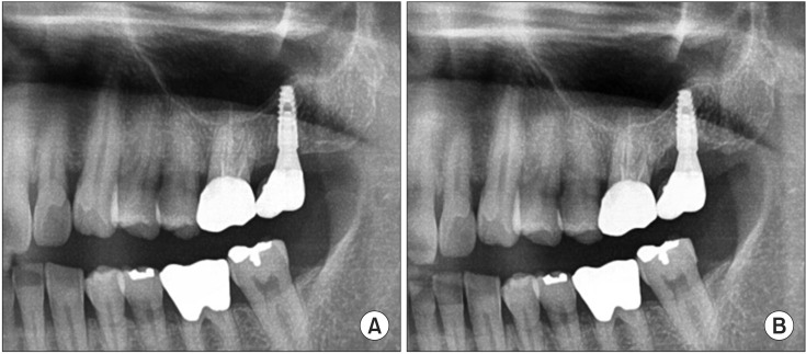
Fig. 1 A. One dental implant was placed at the maxillary left second molar site with an osteotome sinus elevation technique in 2010. B. Marginal bone loss surrounding an implant was observed in group M (bone loss less than 2 mm at the crestal aspect of the implant) in 2015.
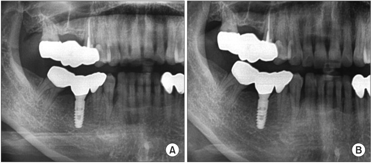
Fig. 2 A. One dental implant was placed at the mandibular right first molar in 2011. B. A 4-year follow-up radiograph demonstrated excellent maintenance of marginal bone surrounding an implant.
Cited by 1 articles
-
Randomized clinical trial to evaluate the efficacy and safety of two types of sandblasted with large-grit and acid-etched surface implants with different surface roughness
Jun-Hyung Jeon, Min-Joong Kim, Pil-Young Yun, Deuk-Won Jo, Young-Kyun Kim
J Korean Assoc Oral Maxillofac Surg. 2022;48(4):225-231. doi: 10.5125/jkaoms.2022.48.4.225.
Reference
-
1. Brånemark PI, Adell R, Albrektsson T, Lekholm U, Lundkvist S, Rockler B. Osseointegrated titanium fixtures in the treatment of edentulousness. Biomaterials. 1983; 4:25–28. PMID: 6838955.
Article2. Albrektsson T, Brånemark PI, Hansson HA, Lindström J. Osseointegrated titanium implants. Requirements for ensuring a longlasting, direct bone-to-implant anchorage in man. Acta Orthop Scand. 1981; 52:155–170. PMID: 7246093.3. Wennerberg A, Ektessabi A, Albrektsson T, Johansson C, Andersson B. A 1-year follow-up of implants of differing surface roughness placed in rabbit bone. Int J Oral Maxillofac Implants. 1997; 12:486–494. PMID: 9274077.4. Trisi P, Lazzara R, Rao W, Rebaudi A. Bone-implant contact and bone quality: evaluation of expected and actual bone contact on machined and osseotite implant surfaces. Int J Periodontics Restorative Dent. 2002; 22:535–545. PMID: 12516825.5. Piattelli M, Scarano A, Paolantonio M, Iezzi G, Petrone G, Piattelli A. Bone response to machined and resorbable blast material titanium implants: an experimental study in rabbits. J Oral Implantol. 2002; 28:2–8. PMID: 12498456.
Article6. Block MS, Kent JN, Kay JF. Evaluation of hydroxylapatite-coated titanium dental implants in dogs. J Oral Maxillofac Surg. 1987; 45:601–607. PMID: 3037051.
Article7. Wennerberg A, Albrektsson T. On implant surfaces: a review of current knowledge and opinions. Int J Oral Maxillofac Implants. 2010; 25:63–74. PMID: 20209188.8. Cochran DL, Buser D, ten Bruggenkate CM, Weingart D, Taylor TM, Bernard JP, et al. The use of reduced healing times on ITI implants with a sandblasted and acid-etched (SLA) surface: early results from clinical trials on ITI SLA implants. Clin Oral Implants Res. 2002; 13:144–153. PMID: 11952734.9. Buser D, Janner SF, Wittneben JG, Brägger U, Ramseier CA, Salvi GE. 10-year survival and success rates of 511 titanium implants with a sandblasted and acid-etched surface: a retrospective study in 303 partially edentulous patients. Clin Implant Dent Relat Res. 2012; 14:839–851. PMID: 22897683.
Article10. Romanos GE, Nentwig GH. Single molar replacement with a progressive thread design implant system: a retrospective clinical report. Int J Oral Maxillofac Implants. 2000; 15:831–836. PMID: 11151582.11. Bowers KT, Keller JC, Randolph BA, Wick DG, Michaels CM. Optimization of surface micromorphology for enhanced osteoblast responses in vitro. Int J Oral Maxillofac Implants. 1992; 7:302–310. PMID: 1289255.12. Rich A, Harris AK. Anomalous preferences of cultured macrophages for hydrophobic and roughened substrata. J Cell Sci. 1981; 50:1–7. PMID: 7033247.
Article13. Martin JY, Schwartz Z, Hummert TW, Schraub DM, Simpson J, Lankford J Jr, et al. Effect of titanium surface roughness on proliferation, differentiation, and protein synthesis of human osteoblastlike cells (MG63). J Biomed Mater Res. 1995; 29:389–401. PMID: 7542245.
Article14. den Braber ET, de Ruijter JE, Smits HT, Ginsel LA, von Recum AF, Jansen JA. Effect of parallel surface microgrooves and surface energy on cell growth. J Biomed Mater Res. 1995; 29:511–518. PMID: 7622536.
Article15. Wennerberg A, Albrektsson T, Andersson B, Krol JJ. A histomorphometric and removal torque study of screw-shaped titanium implants with three different surface topographies. Clin Oral Implants Res. 1995; 6:24–30. PMID: 7669864.16. Wennerberg A, Albrektsson T, Lausmaa J. Torque and histomorphometric evaluation of c.p. titanium screws blasted with 25- and 75-microns-sized particles of Al2O3. J Biomed Mater Res. 1996; 30:251–260. PMID: 9019491.17. Im JH, Kim SG, Oh JS, Lim SC. A comparative study of stability after the installation of 2 different surface types of implants in the maxillae of dogs. Implant Dent. 2015; 24:586–591. PMID: 26076390.
Article18. Elkhaweldi A, Lee DH, Wang W, Cho SC. The survial rate of RBM surface versus SLA surface in geometrically identical implant design. J Oral Bio. 2014; 1:1–8.19. Higuchi KW, Folmer T, Kultje C. Implant survival rates in partially edentulous patients: a 3-year prospective multicenter study. J Oral Maxillofac Surg. 1995; 53:264–268. PMID: 7861276.20. Quirynen M, Bollen CM, Eyssen H, van Steenberghe D. Microbial penetration along the implant components of the Brånemark system. An in vitro study. Clin Oral Implants Res. 1994; 5:239–244. PMID: 7640338.
Article21. Albrektsson T, Zarb G, Worthington P, Eriksson AR. The long-term efficacy of currently used dental implants: a review and proposed criteria of success. Int J Oral Maxillofac Implants. 1986; 1:11–25. PMID: 3527955.22. Lazzara RJ, Porter SS. Platform switching: a new concept in implant dentistry for controlling postrestorative crestal bone levels. Int J Periodontics Restorative Dent. 2006; 26:9–17. PMID: 16515092.
- Full Text Links
-
- Actions
-
Cited
- CITED
-
- Close
- Share
-
- Similar articles
-
- Comparative Study of the Early Loading of Resorbable Blasting Media and Sandblasting with Large-grit and Acid-etching Surface Implants: A Retrospective Cohort Study
- Evaluation of Dental Implant Stability with or without Photoactivated Surface Treatment
- Effect of surface modification on bond strength in titanium-porcelain system
- A shear bond strength of resin cements bonded to pressable porcelain with various surface treatments
- Scanning Electron Microscopic Study of the Effects of Citric Acid on the Change of Implant Surface According to Application Time
