J Korean Soc Radiol.
2015 Dec;73(6):424-427. 10.3348/jksr.2015.73.6.424.
MRI Induced Second-Degree Burn in a Patient with Extremely Large Uterine Leiomyomas: A Case Report
- Affiliations
-
- 1Department of Radiology, Hanyang University Medical Center, Hanyang University College of Medicine, Seoul, Korea. msbbogri@naver.com
- 2Division of Gynecologic Oncology and Gynecologic Minimally Invasive Surgery, Department of Obstetrics and Gynecology, Hanyang University Medical Center, Hanyang University College of Medicine, Seoul, Korea.
- KMID: 2130951
- DOI: http://doi.org/10.3348/jksr.2015.73.6.424
Abstract
- Burns and thermal injuries related with magnetic resonance imaging (MRI) are rare. Previous literature indicates that medical devices with cable, cosmetics or tattoo are known as risk factors for burns and thermal injuries. However, there is no report of MRI-related burns in Korea. Herein, we reported a case of deep second degree burn after MRI in a 38-year-old female patient with multiple uterine leiomyomas including some that were large and degenerated. The large uterine leiomyoma-induced protruded anterior abdominal wall in direct contact with the body coil during MRI was suspected as the cause of injury, by retrospective analysis. Therefore, awareness of MRI related thermal injury is necessary to prevent this hazard, together with extreme care during MRI.
MeSH Terms
Figure
-
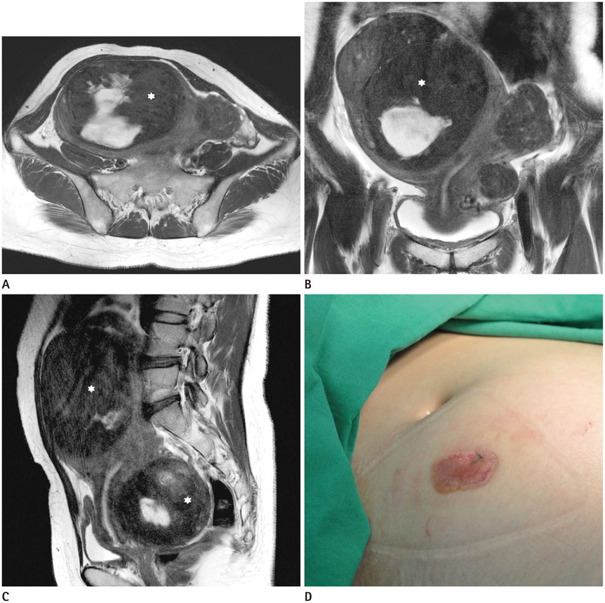
Fig. 1 A 38-year-old female patient with multiple uterine leiomyomas including one in the right anterior that was extremely large. A-C. T2-weighted axial (A) and coronal (B), and sagittal (C) images show 2 extremely large degenerated subserosal type leiomyomas in the right anterior and posterior side of the uterus (asterisks) and other multiple T2-hypointense leiomyomas. The adjacent right lower anterior abdominal wall is protruded by the mass effect of the large leiomyoma. D. Clinical photograph of the same patient. A deep 2nd degree burn is noted at the anterior abdominal wall of right lower abdomen that was protruded by the large uterine leiomyoma.
Reference
-
1. Formica D, Silvestri S. Biological effects of exposure to magnetic resonance imaging: an overview. Biomed Eng Online. 2004; 3:11.2. Knopp MV, Essig M, Debus J, Zabel HJ, van Kaick G. Unusual burns of the lower extremities caused by a closed conducting loop in a patient at MR imaging. Radiology. 1996; 200:572–575.3. Masaki F, Shuhei Y, Riko K, Yohjiro M. Iatrogenic second-degree burn caused by a catheter encased tubular braid of stainless steel during MRI. Burns. 2007; 33:1077–1079.4. Shellock FG, Crues JV. MR procedures: biologic effects, safety, and patient care. Radiology. 2004; 232:635–652.5. Franiel T, Schmidt S, Klingebiel R. First-degree burns on MRI due to nonferrous tattoos. AJR Am J Roentgenol. 2006; 187:W556.6. Haik J, Daniel S, Tessone A, Orenstein A, Winkler E. MRI induced fourth-degree burn in an extremity, leading to amputation. Burns. 2009; 35:294–296.7. Pietryga JA, Fonder MA, Rogg JM, North DL, Bercovitch LG. Invisible metallic microfiber in clothing presents unrecognized MRI risk for cutaneous burn. AJNR Am J Neuroradiol. 2013; 34:E47–E50.8. Dempsey MF, Condon B. Thermal injuries associated with MRI. Clin Radiol. 2001; 56:457–465.9. Landman A, Goldfarb S. Magnetic resonance-induced thermal burn. Ann Emerg Med. 2008; 52:308–309.
- Full Text Links
-
- Actions
-
Cited
- CITED
-
- Close
- Share
-
- Similar articles
-
- Deep Burn Injuries on the Lower Abdomen after HIFU Treatment for Uterine Myoma
- Skin Burn after Laparoscopic Radiofrequency Thermal Ablation for Uterine Myoma : A case report
- A Case of Crushing Burn Injury: A Case Report
- Magnetic Resonance Guided Focused Ultrasound Induced Dermal Burn, Case Report
- Current Medical Therapy for Uterine Leiomyomas
