Breast Metastasis from Gastric Adenocarcinoma Mimicking Normal Breast Parenchyma on Ultrasound: A Case Report
- Affiliations
-
- 1Department of Radiology, Dong-A University Hospital, Busan, Korea. jhrad@dau.ac.kr
- 2Department of Radiology, Inje University Busan Paik Hospital, Busan, Korea.
- 3Department of Pathology, Dong-A University Hospital, Busan, Korea.
- 4Department of Surgery, Dong-A University Hospital, Busan, Korea.
- KMID: 2130945
- DOI: http://doi.org/10.3348/jksr.2015.73.6.393
Abstract
- Breast metastases from extramammary malignancies are uncommon. Although metastatic lesions show variable radiologic features, there have been few reports of metastatic breast cancer with negative sonographic findings. Furthermore, the results of several studies have indicated a high negative predictive value when ultrasonographic and mammographic findings were normal in the setting of a palpable lump, and follow-up is recommended when the physical examination is not highly suspicious. Herein, we report a case of a 26-year-old woman with breast metastasis from a known gastric adenocarcinoma, which had negative findings without any evidence of suspicious features for malignancy on the initial mammogram and ultrasound.
MeSH Terms
Figure
-
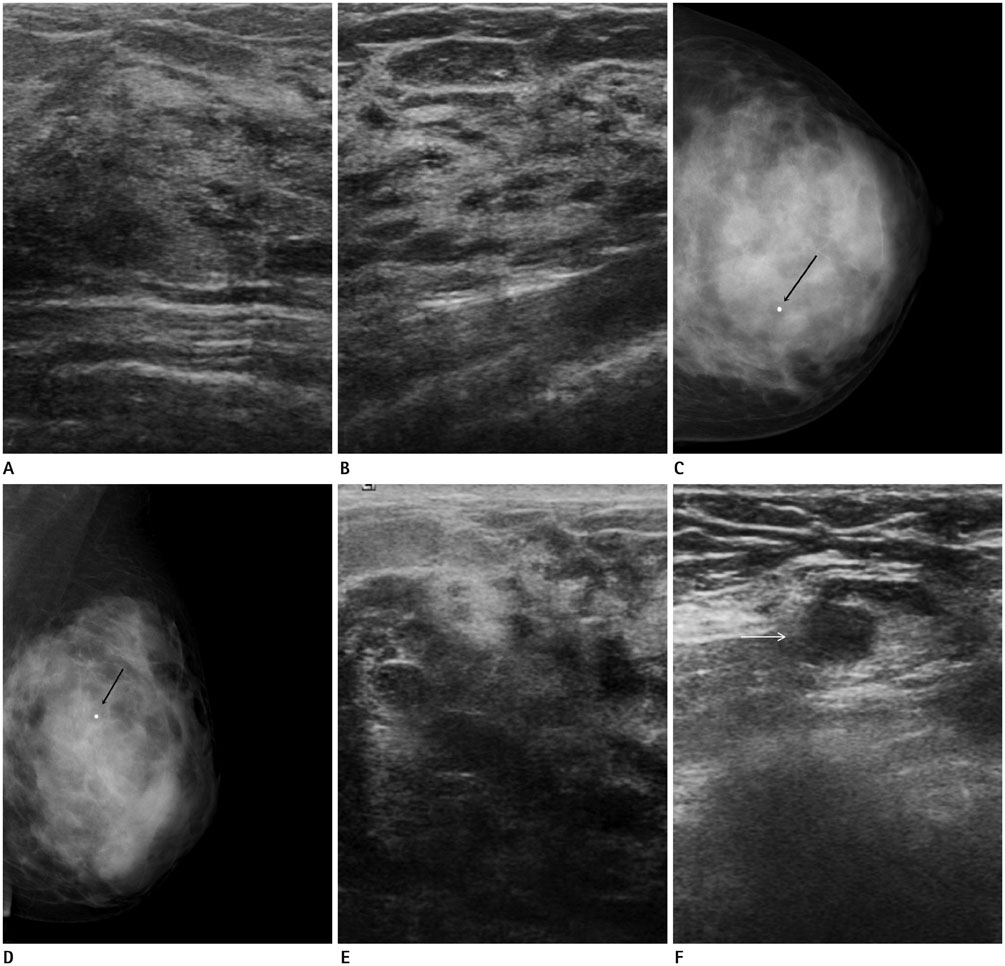
Fig. 1 A 26-year-old female with a palpable lump in the left breast. A. Initial ultrasonographic (US) of the palpable area at the left 10-o'clock position shows heterogeneous fibroglandular tissue without a discrete mass lesion. B. Symmetric parenchymal echotexture is noted in the corresponding area of the contralateral breast (right 2-o'clock). C, D. Left craniocaudal (C) and mediolateral oblique (D) mammograms show extremely dense parenchyma and no abnormal findings at the palpable radiopaque BB marker area (black arrows) of the left-upper inner breast. E. Follow-up US after 1 month was conducted due to an enlargement of the palpable breast lump. Targeted US shows a further heterogeneous echotexture characterized by large areas of decreased echogenecity without a discrete mass lesion. Skin thickening is also observed. F. US of the ipsilateral axillary lymph node shows eccentric cortical thickening (white arrow).

Fig. 2 Histopathology of the breast biopsy specimen. A. The tumor cells seem to be signet ring cells with abundant intracytoplasmic mucin (arrows), and poorly differentiated small pleomorphic features are frequently recognized as well (hematoxylin and eosin, × 200). B, C. Cytokeratin (CK) 20 (B) and CK7 (C) staining are both positive (× 400).
Reference
-
1. Wood B, Sterrett G, Frost F, Swarbrick N. Diagnosis of extramammary malignancy metastatic to the breast by fine needle biopsy. Pathology. 2008; 40:345–351.2. Surov A, Fiedler E, Holzhausen HJ, Ruschke K, Schmoll HJ, Spielmann RP. Metastases to the breast from non-mammary malignancies: primary tumors, prevalence, clinical signs, and radiological features. Acad Radiol. 2011; 18:565–574.3. Shetty MK, Shah YP. Prospective evaluation of the value of negative sonographic and mammographic findings in patients with palpable abnormalities of the breast. J Ultrasound Med. 2002; 21:1211–1216. quiz 1217-12194. Dennis MA, Parker SH, Klaus AJ, Stavros AT, Kaske TI, Clark SB. Breast biopsy avoidance: the value of normal mammograms and normal sonograms in the setting of a palpable lump. Radiology. 2001; 219:186–191.5. Lee JH, Kim EK, Yoon SK, Choi S, Nam KJ, Cho SH, et al. The Clinical Significance of Normal Mammograms and Normal Sonograms in Patients with Palpable Abnormalities of the Breast. J Korean Radiol Soc. 2006; 55:299–304.6. Mun SH, Ko EY, Han BK, Shin JH, Kim SJ, Cho EY. Breast metastases from extramammary malignancies: typical and atypical ultrasound features. Korean J Radiol. 2014; 15:20–28.7. Abbas J, Wienke A, Spielmann RP, Bach AG, Surov A. Intramammary metastases: comparison of mammographic and ultrasound features. Eur J Radiol. 2013; 82:1423–1430.8. Ahn SJ, Kim SK, Kim EK. Metastatic breast cancer from rhabdomyosarcoma mimicking normal breast parenchyma on sonography. J Ultrasound Med. 2010; 29:489–492.9. Kyoung Jung H, Kim EK, Yun M, Jung Kim M, Young Kwak J. Bilateral breasts involvement in Burkitt's lymphoma detected only by FDG-PET. Clin Imaging. 2006; 30:57–59.10. American College of Radiology. ACR BI-RADS Mammography. In : Sickles EA, D'Orsi CJ, Bassett LW, Appleton CM, Berg WA, Burnside ES, editors. ACR BI-RADS® Atlas, Breast Imaging Reporting and Data System. Reston, VA: American College of Radiology;2013.
- Full Text Links
-
- Actions
-
Cited
- CITED
-
- Close
- Share
-
- Similar articles
-
- Sparganosis of the Breast that Mimicked Metastasis: A Case Report
- An Unusual Case of Gastric Cancer Presenting with Breast Metastasis with Pleomorphic Microcalcifications
- A case of stomach metastasis from breast cancer
- Adenoid Cystic Carcinoma of the Breast: A Case Report
- Synchronously Diagnosed Gastric Metastasis from Invasive Lobular Breast Carcinoma, Mimicking Primary Gastric Carcinoma
