Change of Distribution and Timing of Bite Force after Botulinum Toxin Type A Injection Evaluated by a Computerized Occlusion Analysis System
- Affiliations
-
- 1Department of Orofacial Pain and Oral Medicine, College of Dentistry, Yonsei University, Seoul, Korea. hjahn@yuhs.ac
- KMID: 2130842
- DOI: http://doi.org/10.3349/ymj.2014.55.4.1123
Abstract
- PURPOSE
The aim of this study was to determine the force distribution and pattern of mastication after injection of botulinum toxin type A (BTX-A) into both masseter muscles. The hypothesis to be tested was that the difference between right and left balance of occlusal force diminishes over time following BTX-A injection.
MATERIALS AND METHODS
Fifteen patients were submitted to BTX-A injection therapy for subjective masseter hypertrophy. A total of 25 U of BTX-A (50 U in total) was injected into two points located 1 cm apart at the center of the lower one-third of both masseter muscles. All patients were examined using the T-Scan occlusion analysis system before and 4, 8, 12, and 24 weeks after BTX-A injection.
RESULTS
A significant change in force balance was found between the right and left sides over time and the difference between the two sides decreased with the time post-injection, reaching a minimum at 12 weeks. Comparison of the force balance between the anterior and posterior occlusions revealed no significant difference at any of the time points. The occlusion and disclusion times (right and left sides) did not differ significantly with time since BTX-A injection.
CONCLUSION
A decline in the difference in the clenching force between the left and right sides was found with increasing time up to 12 weeks following BTX-A injection.
Keyword
MeSH Terms
Figure
-
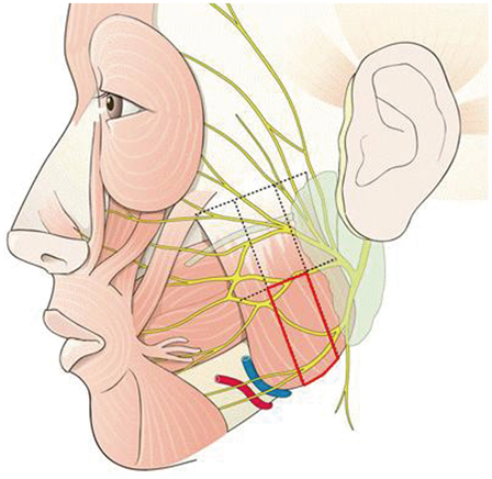
Fig. 1 The red boxed area shows the botulinum toxin type A injection site.
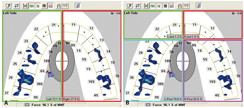
Fig. 2 Balance of clenching force in a T-Scan movie (A: divided into two parts: left and right sides, B: divided into four parts labeled in clockwise direction: anterior left, anterior right, posterior right, posterior left).

Fig. 3 (A) Occlusion time. (B) Disclusion time. (A) A1 is the point the teeth start to occlude, B1 is the point the dentition is fully occluded, and the difference of A1 and B1 is occlusion time, marked as OT. (B) C1 is the point lateral movement starts, D1 is the point the dentition is fully separated and the difference is disclusion time, marked as DT. OT, occlusion time; DT, disclusion time.
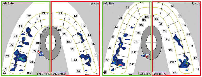
Fig. 4 Force distribution on right and left sides before BTX-A injection (A) and 12 weeks thereafter (B). (A) In the left figure, before botulinum toxin injection the distribution of the left and right force was uneven, 72.1% and 27.9%. 12 weeks post injection, (B) the right figure, the distribution evened as left 58.1% and right 27.9%. BTX-A, botulinum toxin type A.
Reference
-
1. Clark GT. The management of oromandibular motor disorders and facial spasms with injections of botulinum toxin. Phys Med Rehabil Clin N Am. 2003; 14:727–748.
Article2. Freund B, Schwartz M, Symington JM. Botulinum toxin: new treatment for temporomandibular disorders. Br J Oral Maxillofac Surg. 2000; 38:466–471.
Article3. Lang AM. Botulinum toxin therapy for myofascial pain disorders. Curr Pain Headache Rep. 2002; 6:355–360.
Article4. Lovell BV, Marmura MJ. New therapeutic developments in chronic migraine. Curr Opin Neurol. 2010; 23:254–258.
Article5. Bhogal PS, Hutton A, Monaghan A. A review of the current uses of Botox for dentally-related procedures. Dent Update. 2006; 33:165–168.
Article6. Soboļeva U, Lauriņa L, Slaidiņa A. The masticatory system--an overview. Stomatologija. 2005; 7:77–80.7. Okeson JP. Management of temporomandibular disorders and occlusion. 6th ed. St. Louis, Missouri: Elsevier Mosby Publishers;1993.8. Fallah HM, Currimbhoy S. Use of botulinum toxin A for treatment of myofascial pain and dysfunction. J Oral Maxillofac Surg. 2012; 70:1243–1245.
Article9. Kerstein RB, Lowe M, Harty M, Radke J. A force reproduction analysis of two recording sensors of a computerized occlusal analysis system. Cranio. 2006; 24:15–24.
Article10. Montgomery MW, Shuman L, Morgan A. T-scan dental force analysis for routine dental examination. Dent Today. 2011; 30:112–114. 11611. Hu KS, Kim ST, Hur MS, Park JH, Song WC, Koh KS, et al. Topography of the masseter muscle in relation to treatment with botulinum toxin type A. Oral Surg Oral Med Oral Pathol Oral Radiol Endod. 2010; 110:167–171.
Article12. Kerstein RB. Combining technologies: a computerized occlusal analysis system synchronized with a computerized electromyography system. Cranio. 2004; 22:96–109.
Article13. Makofsky HW, Sexton TR, Diamond DZ, Sexton MT. The effect of head posture on muscle contact position using the T-Scan system of occlusal analysis. Cranio. 1991; 9:316–321.
Article14. Kerstein RB, Wright NR. Electromyographic and computer analyses of patients suffering from chronic myofascial pain-dysfunction syndrome: before and after treatment with immediate complete anterior guidance development. J Prosthet Dent. 1991; 66:677–686.
Article15. Aoki KR, Ranoux D, Wissel J. Using translational medicine to understand clinical differences between botulinum toxin formulations. Eur J Neurol. 2006; 13:Suppl 4. 10–19.
Article16. Addante RR. Masseter muscle hypertrophy: report of case and literature review. J Oral Maxillofac Surg. 1994; 52:1199–1202.
Article17. Rispoli DZ, Camargo PM, Pires JL Jr, Fonseca VR, Mandelli KK, Pereira MA. Benign masseter muscle hypertrophy. Braz J Otorhinolaryngol. 2008; 74:790–793.
Article18. Simpson LL. The origin, structure, and pharmacological activity of botulinum toxin. Pharmacol Rev. 1981; 33:155–188.19. Drachman DB. Atrophy of skeletal muscle in chick embryos treated with botulinum toxin. Science. 1964; 145:719–721.
Article20. Melling J, Hambleton P, Shone CC. Clostridium botulinum toxins: nature and preparation for clinical use. Eye (Lond). 1988; 2(Pt 1):16–23.
Article21. Filippi GM, Errico P, Santarelli R, Bagolini B, Manni E. Botulinum A toxin effects on rat jaw muscle spindles. Acta Otolaryngol. 1993; 113:400–404.
Article22. Holds JB, Alderson K, Fogg SG, Anderson RL. Motor nerve sprouting in human orbicularis muscle after botulinum A injection. Invest Ophthalmol Vis Sci. 1990; 31:964–967.23. Moreno-López B, de la Cruz RR, Pastor AM, Delgado-García JM. Botulinum neurotoxin alters the discharge characteristics of abducens motoneurons in the alert cat. J Neurophysiol. 1994; 72:2041–2044.
Article
- Full Text Links
-
- Actions
-
Cited
- CITED
-
- Close
- Share
-
- Similar articles
-
- The Change of the Maximal Bite-force after Botulinum Toxin A Injection for Lower Face Contouring
- Differences in Psychological Changes after Botulinum Toxin A Administration for Bruxism with Masseter Hypertrophy
- Correction of post-traumatic anterior open bite by injection of botulinum toxin type A into the anterior belly of the digastric muscle: case report
- Influence of Botulinum Toxin A on Corneal Astigmatism
- Electrophysiological Changes after Botulinum Toxin Type A in Children with Cerebral Palsy
