Clear Cell Carcinoma of the Pancreas: A Case Report and Review of the Literature
- Affiliations
-
- 1Department of Internal Medicine, Kangwon National University Hospital, Kangwon National University School of Medicine, Chuncheon, Korea. sysong@kangwon.ac.kr
- 2Department of Pathology, Kangwon National University Hospital, Kangwon National University School of Medicine, Chuncheon, Korea.
Abstract
- Most of the malignant neoplasms of the pancreas demonstrate features that are consistent with adenocarcinoma. According to the WHO classification, primary clear cell carcinoma of the pancreas is rare and it is classified as a "miscellaneous" carcinoma. In addition, there is not an adequate systematic overview that can demonstrate its true existence as a definable entity. We report here on an unusual case of primary pancreatic clear cell carcinoma, which is the first such reported case in Korea. A 66 year old woman presented with abdominal pain and significant weight loss over the previous three weeks. On the abdominal computed tomography (CT), we detected an abdominal mass involving the pancreas tail and liver, and clear cell carcinoma with rhabdoid feature was seen on the histologic evaluation. The tumor cells showed well defined cell membranes, clear cytoplasm and prominent cell boundaries. The immunohistochemical stains showed positive reactions to antibodies against pan-cytokeratin, cytokeratin 7, carcinoembryonic antigen (CEA) and epithelial membrane antigen (EMA). On the other hand, there was a negative reaction for cytokeratin 20, chromogranin, synaptophysin, smooth muscle actin and HMB-45. She was diagnosed with a primary pancreatic clear cell carcinoma with hepatic metastasis and she received palliative gemcitabine chemotherapy. The patient died one month later of pancreatic cancer progression.
Keyword
MeSH Terms
-
Abdominal Pain
Actins
Adenocarcinoma
Antibodies
Carcinoembryonic Antigen
Cell Membrane
Coloring Agents
Cytoplasm
Deoxycytidine
Female
Hand
Humans
Keratin-20
Keratin-7
Korea
Liver
Mucin-1
Muscle, Smooth
Neoplasm Metastasis
Pancreas
Pancreatic Neoplasms
Rhabdoid Tumor
Synaptophysin
Weight Loss
Actins
Antibodies
Carcinoembryonic Antigen
Coloring Agents
Deoxycytidine
Keratin-20
Keratin-7
Mucin-1
Synaptophysin
Figure
-
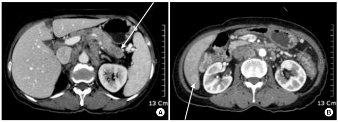
Fig. 1 Abdominal CT scans showed a low attenuated mass in the pancreas tail (A) and another small low attenuated lesion in the right inferior tip of the liver (B).
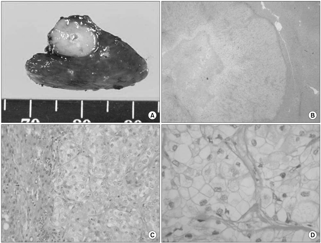
Fig. 2 Gross photograph showing a well circumscribed, firm and grayish white tumor of the liver (A). Microscopic findings of the liver tissue showing round to oval cells with large nuclei, a well defined cell membrane and clear cytoplasm with prominent cell boundaries (H & E, B: ×40, C: ×200, D: ×400).
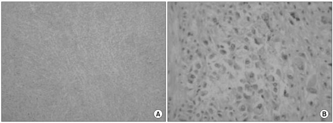
Fig. 3 Microscopic findings of the pancreatic tumor showing the rhabdoid features (H & E, A: ×40, B:×200).
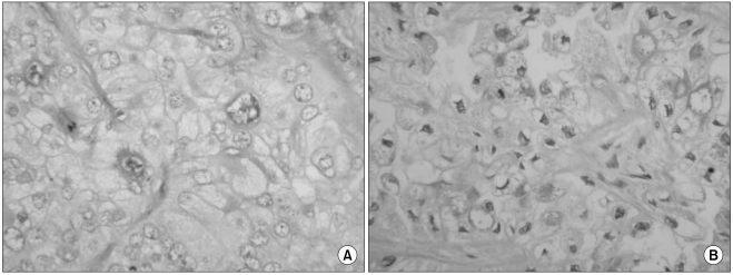
Fig. 4 Histochemical staining of the clear cell carcinoma: the cytoplasm of the clear cells focally stains with PAS (A) and it does not stain with PAS after diastase treatment (B). (×400).
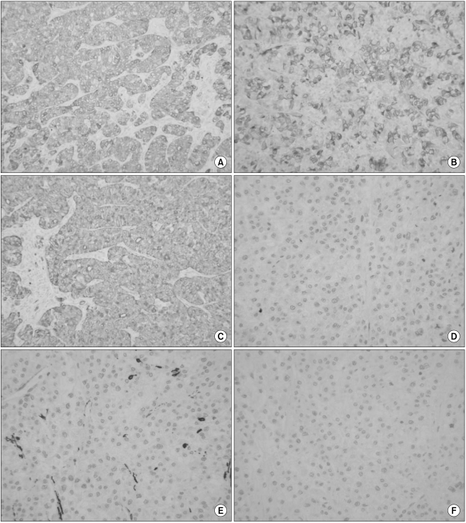
Fig. 5 Immunohistochemical findings. The tumor cells showed positivity for EMA (A, ×200), CEA (B, ×200) and cytokeratin 7 (C, ×200). However, the tumor cells showed negativity for synaptophysin (D, ×200), smooth muscle actin (E, ×200) and HMB-45 (F, ×200).
Reference
-
1. Korean National Statistics Office. Annual Report on the Cause of Death Statistics. 2007. Daejeon: Korean National Statistics Office.2. Zamboni G, Klöppel G. Hamilton SR, Aaltonen LA, editors. Tumours of the exocrine pancreas. Pathology and genetics of tumours of the digestive system. 2000. Lyon: IARC Press;p. 221–250.3. Wick MR, Ritter JH, Humphrey PA, Nappi O. Clear cell neoplasms of the endocrine system and thymus. Semin Diagn Pathol. 1997; 14:183–202. PMID: 9279975.4. Ritter JH, Mills SE, Gaffey MJ, Nappi O, Wick MR. Clear cell tumors of the alimentary tract and abdominal cavity. Semin Diagn Pathol. 1997; 14:213–219. PMID: 9279977.5. Paik SS, Oh YH, Hong EK, Park MH, Lee JD. Clear cell islet cell tumor of the pancreas: an immunohistochemical and ultrastructual study. Korean J Pathol. 1997; 31:162–166.6. Ray S, Lu Z, Rajendiran S. Clear cell ductal adenocarcinoma of pancreas: a case report and review of literature. Arch Pathol Lab Med. 2004; 128:693–696. PMID: 15163226.7. Kanai N, Nagaki S, Tanaka T. Clear cell carcinoma of the pancreas. Acta Pathol Jpn. 1987; 37:1521–1526. PMID: 3687432.
Article8. Lüttges J, Vogel I, Menke M, Henne-Bruns D, Kremer B, Klöppel G. Clear cell carcinoma of the pancreas: an adenocarcinoma with ductal phenotype. Histopathology. 1998; 32:444–448. PMID: 9639120.
Article9. Jamali M, Serra S, Chetty R. Adenosquamous carcinoma of the pancreas with clear cell and rhabdoid components. JOP. 2007; 8:330–334. PMID: 17495363.10. Sasaki A, Ishio T, Bandoh T, Shibata K, Matsumoto T, Aramaki M, et al. Clear cell carcinoma of the pancreas: an adenocarcinoma with unusual phenotype of duct cell origin. J Hepatobiliary Pancreat Surg. 2004; 11:140–144. PMID: 15127279.11. Kim L, Liao J, Zhang M, Talamonti M, Bentrem D, Rao S, et al. Clear cell carcinoma of the pancreas: histopathologic features and a unique biomarker: hepatocyte nuclear factor-1β. Mod Pathol. 2008; 21:1075–1083. PMID: 18536653.
Article12. Weeks DA, Beckwith JB, Mierau GW. Rhabdoid tumor. An entity or a phenotype? Arch Pathol Lab Med. 1989; 113:113–114. PMID: 2916901.
- Full Text Links
-
- Actions
-
Cited
- CITED
-
- Close
- Share
-
- Similar articles
-
- A Case of Primary Small Cell Carcinoma of Pancreas
- A Case of Clear Cell Squamous Cell Carcinoma
- A case of clear cell carcinoma arising from the endometriosis of the paraovarian cyst
- A Case of Clear Cell Basal Cell Carcinoma
- Surgery for metastatic renal cell carcinoma in the pancreatic head: A case report and literature review
