J Korean Ophthalmol Soc.
2010 Sep;51(9):1287-1291. 10.3341/jkos.2010.51.9.1287.
A Case of Corneal Endothelial Damage by 5-Fluorouracil Inflow Into Anterior Chamber After Subconjunctival Injection
- Affiliations
-
- 1Department of Ophthalmology, Ajou University School of Medicine, Suwon, Korea. chrisahn@ajou.ac.kr
- KMID: 2122299
- DOI: http://doi.org/10.3341/jkos.2010.51.9.1287
Abstract
- PURPOSE
To present a case of corneal endothelial damage caused by inflow of 5-FU after subconjunctival 5-FU injection in a patient undergoing trabeculectomy.
CASE SUMMARY
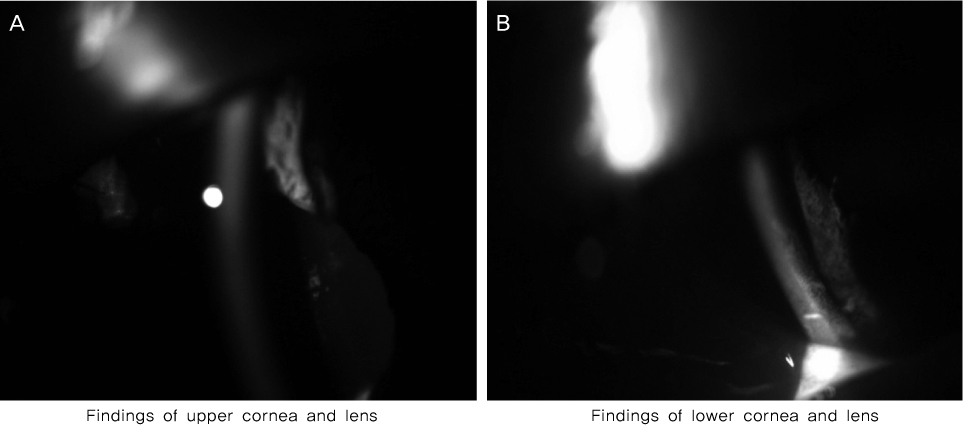
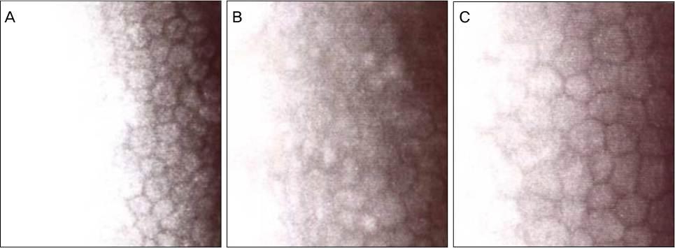
A 65-year-old female patient diagnosed with chronic angle-closure glaucoma underwent trabeculectomy using mitomycin C. Three weeks after the surgery, her intraocular pressure (IOP) elevated, and the follicles had disappeared by trabeculectomy. Subsequently, subconjunctival 5-FU injection was performed. Following the fourth injection, visual acuity abruptly decreased, and corneal edema was observed. Upon presumption of inflow of 5-FU into the anterior chamber, anterior chamber irrigation was performed within 40 minutes. On postoperative day 1, visual acuity decreased from 0.8 to counting fingers, and diffuse corneal edema and anterior capsular opacity of the lens were noted. Three months after the irrigation, visual acuity improved to 0.8. Corneal edema and capsular opacity also improved, however the density of the corneal endothelial count decreased.
CONCLUSIONS
Inflow of 5-FU into the anterior chamber can cause corneal and lenticular toxicity. Anterior chamber irrigation is considered necessary to prevent 5-FU toxicity.
Keyword
MeSH Terms
Figure
Reference
-
1. Allingham RR, Damji KF, Freedman S, et al. Shields' Textbook of Glaucoma. 2005. 5th ed. Philadelphia: Lippincott Williams & Wilkins;577–579.2. Lattanzio FA Jr, Sheppard JD Jr, Allen RC, et al. Do injections of 5-fluorouracil after trabeculectomy have toxic effects on the anterior segment? J Ocul Pharmacol Ther. 2005. 21:223–235.3. Bhermi GS, Holak S, Murdoch IE. Inadvertent exposure of corneal endothelium to 5-fluorouracil. Br J Ophthalmol. 1999. 83:376–377.4. Mazey BJ, Siegel MJ, Siegel LI, Dunn SP. Corneal endothelial toxic effect secondary to fluorouracil needle bleb revision. Arch Ophthalmol. 1994. 112:1411.5. Lama PJ, Fechtner RD. Antifibrotics and wound healing in glaucoma surgery. Surv Ophthalmol. 2003. 48:314–346.6. Nuyts RM, Pels E, Greve EL. The effects of 5-fluorouracil and mitomycin C on the corneal endothelium. Curr Eye Res. 1992. 11:565–570.7. Mannis MJ, Sweet EH, Lewis RA, et al. The effect of fluorouracil on the corneal endothelium. Arch Ophthalmol. 1988. 106:816–817.8. Krachmer JH, Mannis MJ, Holland EJ. Cornea. 2005. Vol. 1:2nd ed. Elsevier;1285–1294.9. Shaarawy TM, Sherwood MB, Hitchings RA, Crowston JG. Glaucoma. Vol. 2 Surgical Management. 2009. Saunders Elsevier;223–229.
- Full Text Links
- Actions
-
Cited
- CITED
-
- Close
- Share
- Similar articles
-
- A Case of Endophthalmitis with Atypical Anterior Symptom by Subconjunctival Dexamethasone Injection after Cataract Surgery
- Filtering Operation with Adjunctive Mitomycin-C in Rabbit: Comparative Study by Subconjunctival Injection and Soaking of Mitomycin-C
- The Comparative Assessment of Filtering Bleb by Timing of subconjunctival Injection of Mitomycin-C in Glaucoma Filtering Surgery
- The Toxic and Morphologic Effects of Mitomycin-C, 5-FU and Genistein on Rabbit Corneal Endothelium
- Prognosis of Ocular Injury Caused by Wasp Sting: Case Reports


