J Korean Ophthalmol Soc.
2015 May;56(5):672-679. 10.3341/jkos.2015.56.5.672.
Comparative Analysis of Clinical Outcome in Penetrating Keratoplasty Using Domestic or Imported Cornea
- Affiliations
-
- 1Department of Ophthalmology, Seoul National University College of Medicine, Seoul, Korea. kmk9@snu.ac.kr
- 2Laboratory of Ocular Regenerative Medicine and Immunology, Seoul Artificial Eye Center, Seoul National University Hospital Biomedical Research Institute, Seoul, Korea.
- 3Department of Ophthalmology, Soonchunhyang University Gumi Hospital, Soonchunhyang University College of Medicine, Gumi, Korea.
- KMID: 2121163
- DOI: http://doi.org/10.3341/jkos.2015.56.5.672
Abstract
- PURPOSE
To compare the survival of corneal grafts and the changes in endothelial cell density in penetrating keratoplasty using domestic or imported corneas.
METHODS
Medical records of 236 eyes of 211 patients who underwent penetrating keratoplasty from November 2004 to August 2011 in Seoul National University Hospital and were followedup at least 1 year were retrospectively reviewed. After excluding the patients who received the combined surgeries with other surgeries except cataract surgery, the eyes were divided into 2 groups depending on the origin of donor tissue resulting in a domestic cornea group (108 eyes) and an imported cornea group (128 eyes). Recipient demographics, preoperative diagnosis, donor age, death-to-preservation time, death-to-operation time and pre-and postoperative visual acuities were compared between the 2 groups. Kaplan-Meier survival and changes in endothelial cell density were analyzed at 3, 6 and 12 months and then every year.
RESULTS
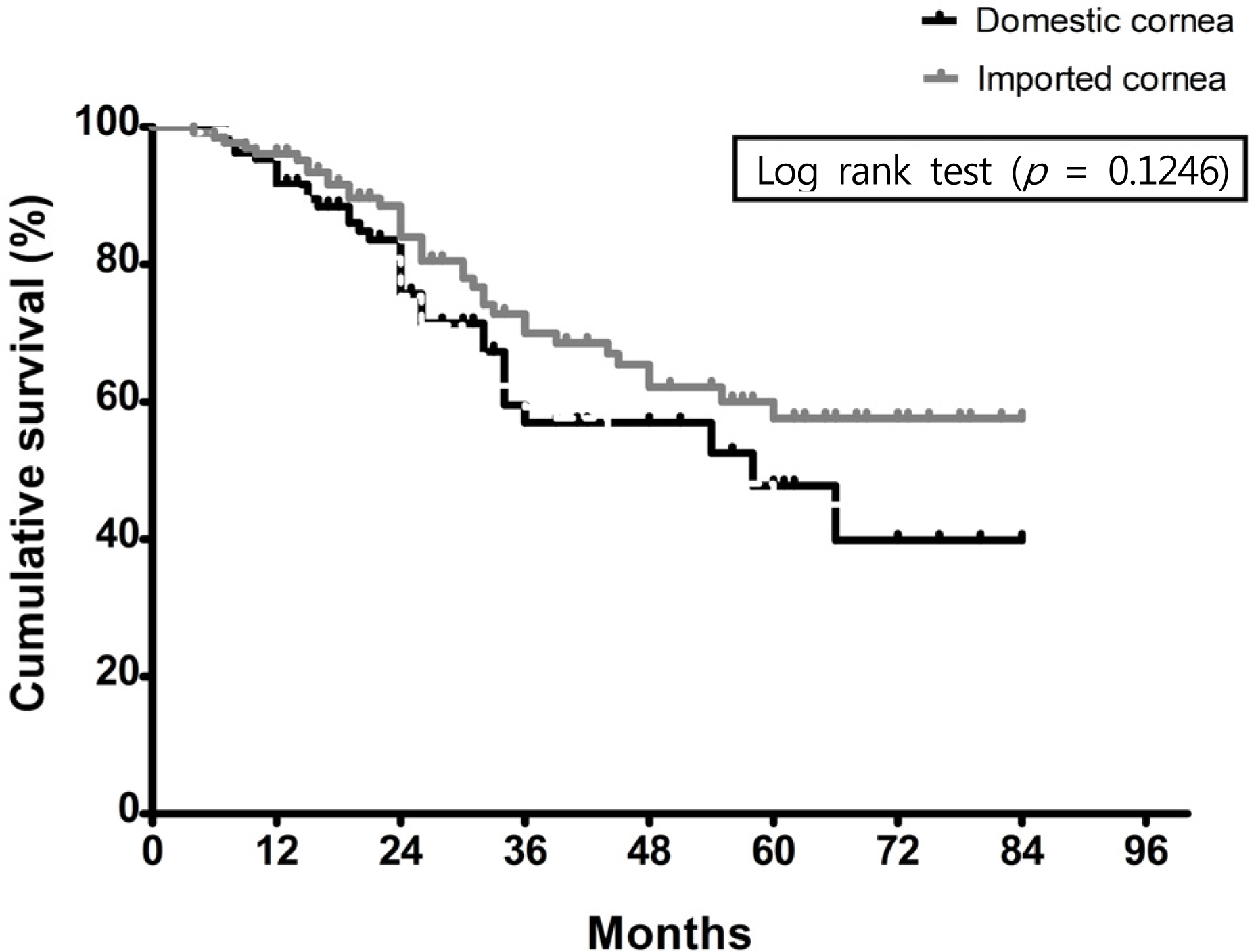
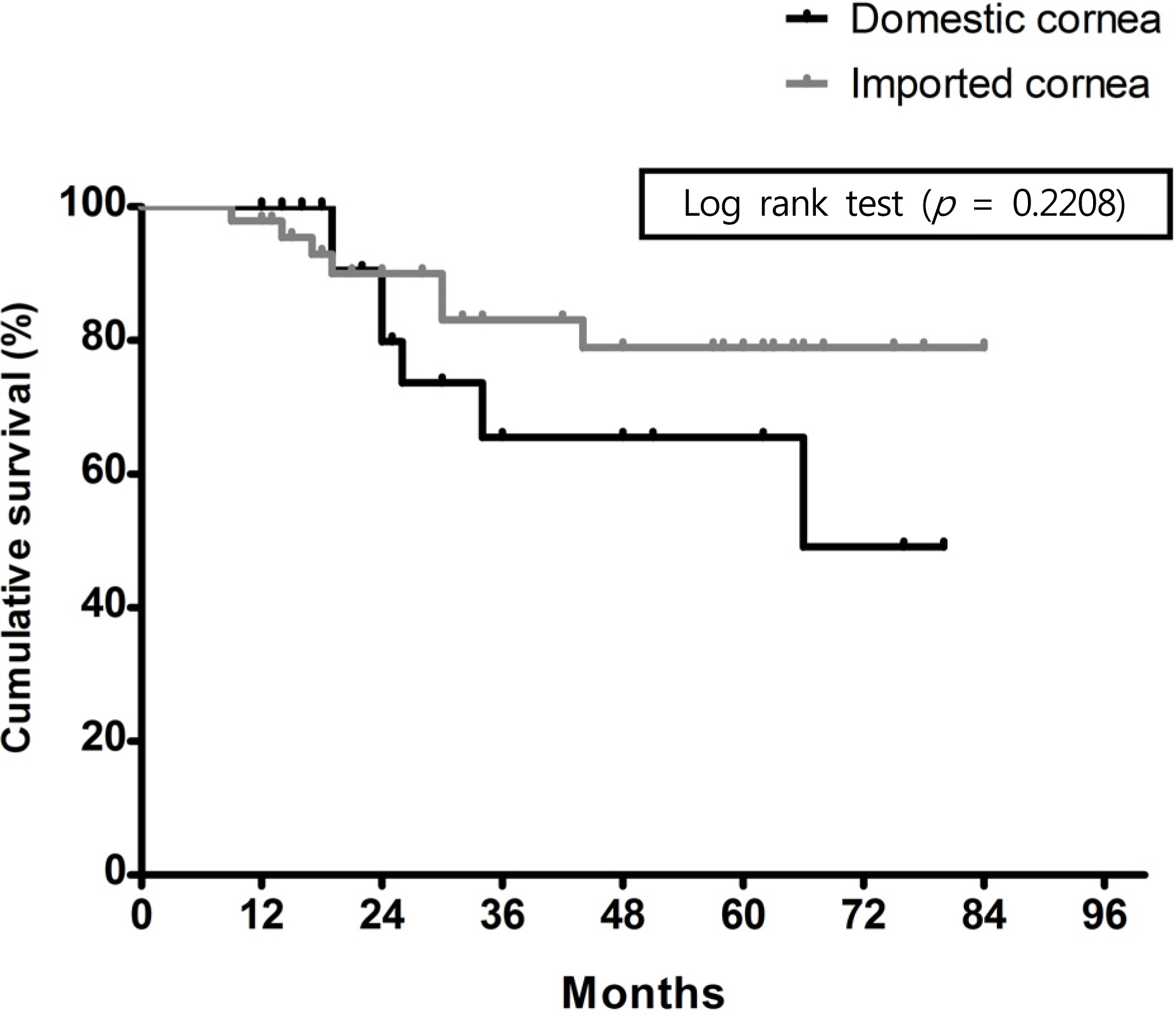
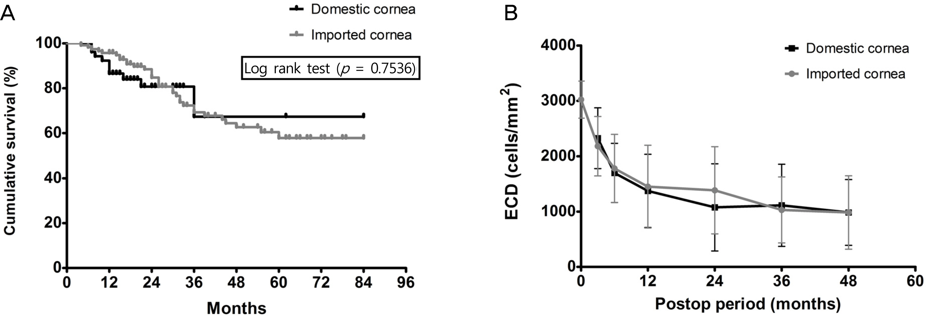
The most common preoperative diagnoses were regraft and corneal opacity in the domestic and imported cornea groups, respectively, without statistical difference. Death-to-preservation time was 8.9 hours and 8.0 hours in the domestic and imported cornea groups, respectively, without statistical difference. However, death-to-operation time was longer in the imported cornea group (4.98 days) than in the domestic cornea group (2.18 days). There were no differences in pre- and postoperative visual acuities, decrease in annual changes in endothelial densities and survival up to 3 years between the 2 groups. In addition, the survival and decreased annual changes in endothelial densities were not different from each other in penetrating keratoplasty combined with cataract surgery or in penetrating keratoplasty for a corneal edema.
CONCLUSIONS
Our study results suggest that clinical outcomes of the penetrating keratoplasty using imported corneas are comparable in efficacy when compared with the clinical outcomes using domestic corneas.
Keyword
MeSH Terms
Figure
Cited by 1 articles
-
Effect of Donor Age on Graft Survival in Primary Penetrating Keratoplasty with Imported Donor Corneas
Hyeon Yoon Kwon, Joon Young Hyon, Hyun Sun Jeon
Korean J Ophthalmol. 2020;34(1):35-45. doi: 10.3341/kjo.2019.0086.
Reference
-
References
1. Hara H, Cooper DK. Xenotransplantation-the future of corneal transplantation? Cornea. 2011; 30:371–8.
Article2. Cho EY, Kim MS. Penetrating keratoplasty before and after estab-lishment of Korean network for organ sharing. J Korean Ophthalmol Soc. 2006; 47:525–30.3. Halberstadt M, Athmann S, Winter R, Hagenah M. Impact of transportation on short-term preserved corneas preserved in Optisol-GS, Likorol, Likorol-DX, and MK-medium. Cornea. 2000; 19:788–91.
Article4. Shimazaki J, Shinozaki N, Shimmura S, et al. Efficacy and safety of international donor sharing: a single-center, case-controlled study on corneal transplantation. Transplantation. 2004; 78:216–20.
Article5. Wagoner MD, Gonnah el-S, Al-Towerki AE; King Khaled Eye Specialist Hospital Cornea Transplant Study Group. Outcome of primary adult optical penetrating keratoplasty with imported donor corneas. Int Ophthalmol. 2010; 30:127–36.
Article6. Park SH, Kim JH, Joo CK. The clinical evaluations of the penetrating keratoplasty with imported donor corneas. J Korean Ophthalmol Soc. 2005; 46:28–34.7. Williams KA, Esterman AJ, Bartlett C, et al. How effective is penetrating corneal transplantation? Factors influencing long-term outcome in multivariate analysis. Transplantation. 2006; 81:896–901.
Article8. Ha D, Kim CK, Lee SE, et al. Penetrating keratoplasty results in 275 cases. J Korean Ophthalmol Soc. 2001; 42:20–9.9. European Eye Bank Association. Technical guidelines for ocular tissue. 2013. Available at: http://www.europeaneyebanks.org/files/Technical_Guidelines_Rev6_Feb2013.pdf.10. Wang IJ, Hu FR. Effect of shaking of corneal endothelial preservation. Curr Eye Res. 1997; 16:1111–8.11. Lee K, Hwang KY, Kim MS. Influence of endothelial cell loss during preservation on graft survival in imported donor cornea. J Korean Ophthalmol Soc. 2013; 54:862–8.
Article12. Yamazoe K, Yamazoe K, Shinozaki N, Shimazaki J. Influence of the precutting and overseas transportation of corneal grafts for Descemet stripping automated endothelial keratoplasty on donor endothelial cell loss. Cornea. 2013; 32:741–4.
Article13. Na YS, Woo SW, Kang JH, Joo MJ. Microbiologic study of imported donor corneas and preserved solutions. J Korean Ophthalmol Soc. 2005; 46:1974–7.14. Hu FR, Tsai AC, Wang IJ, Chang SW. Outcomes of penetrating keratoplasty with imported donor corneas. Cornea. 1999; 18:182–7.
Article15. Armitage WJ, Jones MN, Zambrano I, et al. The suitability of corneas stored by organ culture for penetrating keratoplasty and influence of donor and recipient factors on 5-year graft survival. Invest Ophthalmol Vis Sci. 2014; 55:784–91.
Article16. Kong SJ, Cho K, Kim MS. Analysis of factors affecting the decrease of endothelial cell density in imported donor corneas. J Korean Ophthalmol Soc. 2012; 53:20–6.
Article17. Writing Committee for the Cornea Donor Study Research Group, Mannis MJ, Holland EJ, et al. The effect of donor age on penetrating keratoplasty for endothelial disease: graft survival after 10 years in the Cornea Donor Study. Ophthalmology. 2013; 120:2419–27.
- Full Text Links
- Actions
-
Cited
- CITED
-
- Close
- Share
- Similar articles
-
- Penetrating Keratoplasty before and after Establishment of Korean Network for Organ Sharing
- The Clinical Evaluations of the Penetrating Keratoplasty with Imported Donor Corneas
- Influence of Endothelial Cell Loss During Preservation on Graft Survival in Imported Donor Cornea
- A Case of Anterior Synechiolysis with Lamellar Corneal Dissection in Penetrating Keratoplasty
- Cyclosporin A in High Risk Penetrating Keratoplasty


